Alternate header for print version


Licensing
Attribution Non-Commercial; No Derivatives:This image is licensed under a Creative Commons Attribution, Non-Commercial, No Derivatives License. View License Deed | View Legal Code
tweet  
*CIL – Cell Image Library accession number. Please use this to reference an image.

CIL:41637*  Cite 
Description

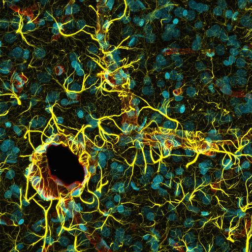
Confocal micrograph of a rat cerebral cortex with astrocytes’ (yellow) endfeet wrapping around blood vessels (red). Cell nuclei are cyan. The image was collected using spectral imaging with 50 Z-slices. Honorable Mention, 2011 Olympus BioScapes Digital Imaging Competition®.

Technical Details

Biological Sources
NCBI Organism Classification
Rattus
Cell Type
astrocyte
Cellular Component
nucleus
astrocyte projection
Biological Context
Biological Process
cerebral cortex development
Mouse Gross Anatomy
blood vessel
Attribution
Names
Madelyn May
Link
2011 Olympus BioScapes Digital Imaging Compet...
OTHER
Rensselaer Polytechnic Institute, Troy, NY, USA
2011 Olympus BioScapes Digital Imaging Competition®
Citation
Digital Object Identifier (DOI)
doi:10.7295/W9CIL41637
Archival Resource Key (ARK)
ark:/b7295/w9cil41637
Imaging
Image Type
recorded image
Image Mode
confocal microscopy
spectral imaging
Parameters Imaged
fluorescence emission
Source of Contrast
distribution of epitope
Processing History
z projection
Dimensions
Spatial Axis Image Size Pixel Size
X 1892px ——
Y 1892px ——