Alternate header for print version


Licensing
Attribution Non-Commercial; No Derivatives:This image is licensed under a Creative Commons Attribution, Non-Commercial, No Derivatives License. View License Deed | View Legal Code
tweet  
*CIL – Cell Image Library accession number. Please use this to reference an image.

CIL:41640*  Cite 
Description

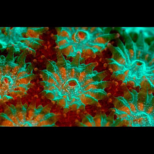
Underwater image of live coral Montastraea annularis. Note polyp tissue (green) around the mouth and base of the tentacles and zooxanthellae or symbiodinium (red fluorescence from chlorophyll) in the tissue between polyps. Tentacles also are visible. Fluorescence microscopy. Honorable Mention, 2011 Olympus BioScapes Digital Imaging Competition®.

Technical Details

Biological Sources
NCBI Organism Classification
Montastraea annularis
symbiodinium
Cell Type
polyps
Cellular Component
chlorophyll
Biological Context
Biological Process
coral colony organization
Attribution
Names
James Nicholson
Link
2011 Olympus BioScapes Digital Imaging Compet...
OTHER
2011 Olympus BioScapes Digital Imaging Competition®
NOAA/NOS/NCCOS Center for Coastal Environmental Health & Biomolecular Research, Fort Johnson Marine Lab, Charleston, SC, USA
Citation
Digital Object Identifier (DOI)
doi:10.7295/W9CIL41640
Archival Resource Key (ARK)
ark:/b7295/w9cil41640
Imaging
Image Type
recorded image
Image Mode
confocal microscopy
Parameters Imaged
fluorescence emission
Source of Contrast
autofluorescence
Sample Preparation
Methods
living tissue
Relation To Intact Cell
whole mounted tissue
Dimensions
Spatial Axis Image Size Pixel Size
X 4072px ——
Y 2636px ——