Alternate header for print version


Licensing
Attribution Non-Commercial Share Alike:This image is licensed under a Creative Commons Attribution, Non-Commercial Share Alike License. View License Deed | View Legal Code
tweet  
*CIL – Cell Image Library accession number. Please use this to reference an image.

CIL:41672*  Cite 
Description

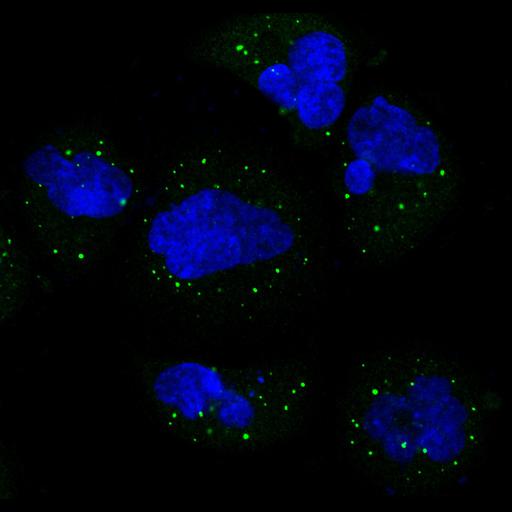
Single slice confocal images representing one optical slice of a cell stained for the Human Protein Atlas (HPA) antibody HPA001619 (shown in green). HPA001619 is a polyclonal anti-EIF4ENIF1 antibody that labels vesicles. The cells are also stained with a reference marker in order to facilitate the annotation of the subcellular distribution of the protein targeted by the HPA antibody. The following probe was used as reference; DAPI for the nucleus (blue).

Technical Details

The use of data and images from this site in publications and presentations is permitted provided that the following conditions are met: The publication and/or presentation are solely for informational and non-commercial purposes. The source of the data and/or image is referred to this site (www.proteinatlas.org) and/or the above publication is cited.

Biological Sources
NCBI Organism Classification
Homo sapiens
Cell Type
epidermoid carcinoma
Cell Line
A-431
Cellular Component
vesicle
nucleus
Biological Context
Biological Process
organelle organization
cytoskeleton organization
Attribution
Names
M Uhlen
P Oksvold
L Fagerberg
E Lundberg
K Jonasson
M Forsberg
M Zwahlen
C Kampf
K Wester
S Hober
H Wernerus
L Björling
F Ponten
Published
Nat Biotechnol. 2010 28(12):1248-50.
Pubmed
21139605
Link
Human Protein Atlas
Citation
Digital Object Identifier (DOI)
doi:10.7295/W9CIL41672
Archival Resource Key (ARK)
ark:/b7295/w9cil41672
Grouping This image is part of a group.
Imaging
Image Type
recorded image
Image Mode
confocal microscopy
Parameters Imaged
fluorescence emission
Source of Contrast
distribution of epitope
Visualization Methods
primary antibody plus labeled secondary antibody
Sample Preparation
Methods
chemically fixed tissue
permeabilized tissue
Relation To Intact Cell
dispersed cells in vitro
Dimensions
Spatial Axis Image Size Pixel Size
X 1254px ——
Y 1191px ——