Alternate header for print version


Licensing
Attribution Non-Commercial; No Derivatives:This image is licensed under a Creative Commons Attribution, Non-Commercial, No Derivatives License. View License Deed | View Legal Code
tweet  
*CIL – Cell Image Library accession number. Please use this to reference an image.

CIL:41733*  Cite 
Description

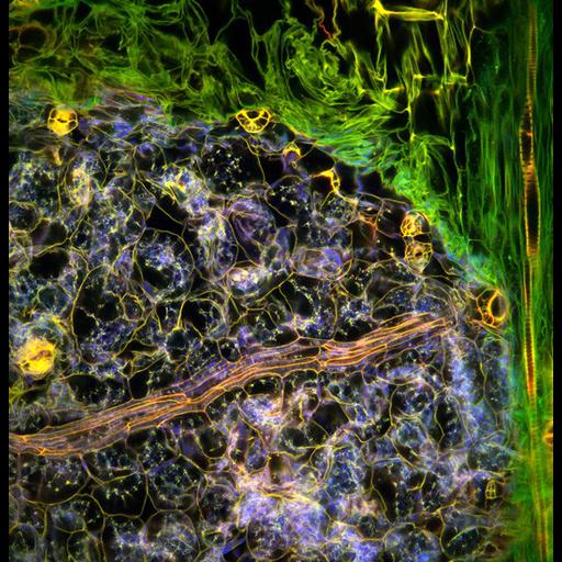
Confocal micrograph showing the tissue structures within the leaf of an Arabidopsis thaliana seedling. The sample was fixed and stained with propidium iodide, which labels DNA, and imaged four years later. Over time, oxidation of the stain differentiates the fluorescent properties in different parts of the tissue so that they can be excited with distinct wavelengths of light from a confocal microscope. The researchers are using these techniques to investigate cellular architecture in plants and gene activity. The horizontal field width of this image is 200 microns. Wellcome Image Award 2012.

Technical Details

B0008269. 2011 Collection: Wellcome Images Copyrighted work available under Creative Commons by-nc-nd 2.0 UK: England & Wales, see http://images.wellcome.ac.uk/indexplus/page/Prices.html

Biological Sources
NCBI Organism Classification
Arabidopsis thaliana
Biological Context
Biological Process
cellular architecture
Plant Growth
leaf development stages
Attribution
Names
Fernan Federici
Jim Haseloff
Link
Wellcome Images
Citation
Digital Object Identifier (DOI)
doi:10.7295/W9CIL41733
Archival Resource Key (ARK)
ark:/b7295/w9cil41733
Imaging
Image Type
recorded image
Image Mode
confocal microscopy
Parameters Imaged
fluorescence emission
Source of Contrast
differences in adsorption or binding of stain
oxidation
Visualization Methods
propidium iodide
Sample Preparation
Methods
chemically fixed tissue
Dimensions
Spatial Axis Image Size Pixel Size
X 533px ——
Y 550px ——