Alternate header for print version


Licensing
Attribution Non-Commercial; No Derivatives:This image is licensed under a Creative Commons Attribution, Non-Commercial, No Derivatives License. View License Deed | View Legal Code
tweet  
*CIL – Cell Image Library accession number. Please use this to reference an image.

CIL:41738*  Cite 
Description

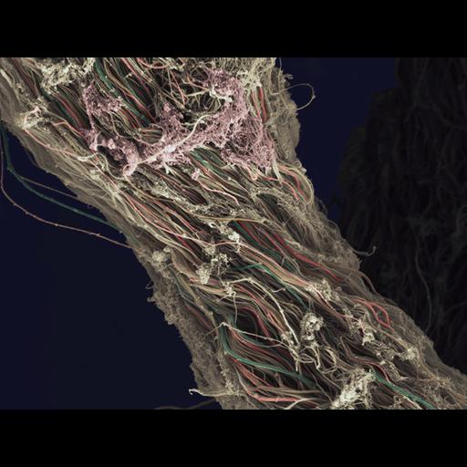
Colorized scanning electron micrograph of collagen/connective tissue removed from a human knee during arthroscopic surgery. The horizontal field width of the image is 16 microns. Wellcome Image Award 2012.

Technical Details

B0008288. 2011 Collection: Wellcome Images Copyrighted work available under Creative Commons by-nc-nd 2.0 UK: England & Wales, see http://images.wellcome.ac.uk/indexplus/page/Prices.html

Biological Sources
NCBI Organism Classification
Homo sapiens
Cellular Component
fibrillar collagen
Biological Context
Biological Process
connective tissue organization
Human Development Anatomy
knee
Attribution
Names
Anne Weston
Link
Wellcome Images
OTHER
London Research Institute, Cancer Center UK
Citation
Digital Object Identifier (DOI)
doi:10.7295/W9CIL41738
Archival Resource Key (ARK)
ark:/b7295/w9cil41738
Imaging
Image Type
recorded image
Image Mode
scanning electron microscopy (SEM)
Parameters Imaged
elastic scattering of electrons
Source of Contrast
differences in deposition of metal shadow
Processing History
colorized
Sample Preparation
Relation To Intact Cell
whole mounted tissue
Dimensions
Spatial Axis Image Size Pixel Size
X 729px ——
Y 550px ——