Alternate header for print version


Licensing
Attribution Non-Commercial; No Derivatives:This image is licensed under a Creative Commons Attribution, Non-Commercial, No Derivatives License. View License Deed | View Legal Code
tweet  
*CIL – Cell Image Library accession number. Please use this to reference an image.

CIL:41833*  Cite 
Description

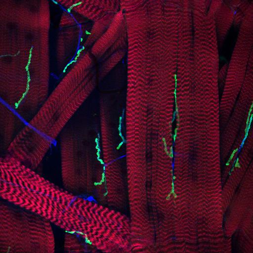
Confocal micrograph showing the complex connectivity at the neuromuscular junction of Drosophila (fruit fly). Muscle fibers shown in red and nerve fibers into neruomuscular junction shown in blue and green. Honorable Mention, 2010 Olympus BioScapes Digital Imaging Competition®.

Technical Details

Biological Sources
NCBI Organism Classification
Drosophila melanogaster
Cell Type
muscle cell
neuron
Cellular Component
neuromuscular junction
Biological Context
Biological Process
neuromuscular junction development
Attribution
Names
Timothy Mosca
2010 Olympus BioScapes Digital Imaging Competition®
Link
2010 Olympus BioScapes Digital Imaging Compe...
OTHER
http://www.olympusbioscapes.com/staticgallery/2010/hm34.html;;;2010 Olympus BioScapes Digital Imaging Competition®
Citation
Digital Object Identifier (DOI)
doi:10.7295/W9CIL41833
Archival Resource Key (ARK)
ark:/b7295/w9cil41833
Imaging
Image Type
recorded image
Image Mode
confocal microscopy
Parameters Imaged
fluorescence emission
Source of Contrast
distribution of a specific protein
Sample Preparation
Relation To Intact Cell
whole mounted tissue
Dimensions
Spatial Axis Image Size Pixel Size
X 1600px ——
Y 1600px ——