Alternate header for print version


Licensing
Attribution Non-Commercial; No Derivatives:This image is licensed under a Creative Commons Attribution, Non-Commercial, No Derivatives License. View License Deed | View Legal Code
tweet  
*CIL – Cell Image Library accession number. Please use this to reference an image.

CIL:42509*  Cite 
Description

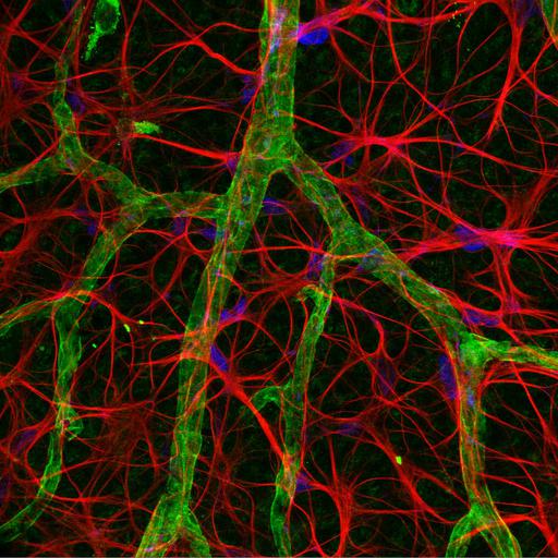
Blood vessels and astrocytes in aging rat retina, confocal imaging, 40x. Blood vessels are shown in blue; astrocytes (supportive cells of the nervous system) are mostly in red. As organisms age, changes in astrocytes might contribute to disease and degeneration. First Prize, 2005 Olympus BioScapes Competition.

Technical Details

Biological Sources
NCBI Organism Classification
Rattus
Cell Type
astrocyte
Cellular Component
astrocyte projection
Biological Context
Biological Process
retinal aging
Mouse Gross Anatomy
retinal blood vessel
Attribution
Names
Hussein Mansour
2005 Olympus BioScapes Competition
Link
2005 Olympus BioScapes Competition
OTHER
University of Sydney, Australia
Citation
Digital Object Identifier (DOI)
doi:10.7295/W9CIL42509
Archival Resource Key (ARK)
ark:/b7295/w9cil42509
Imaging
Image Type
recorded image
Image Mode
confocal microscopy
Parameters Imaged
fluorescence emission
Source of Contrast
distribution of epitope
Sample Preparation
Relation To Intact Cell
whole mounted tissue
Dimensions
Spatial Axis Image Size Pixel Size
X 1024px ——
Y 1024px ——