Alternate header for print version


Licensing
Attribution Non-Commercial; No Derivatives:This image is licensed under a Creative Commons Attribution, Non-Commercial, No Derivatives License. View License Deed | View Legal Code
tweet  
*CIL – Cell Image Library accession number. Please use this to reference an image.

CIL:42515*  Cite 
Description

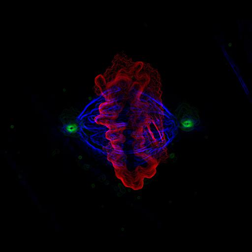
Fluorescence micrograph of a human epithelial cell in mitosis, labeled for alpha tubulin (blue), gamma tubulin (green) and DNA (red). The image was taken with a 100x objective and processed with deconvolution. Honorable Mention, 2004 Olympus BioScapes Competition.

Technical Details

Biological Sources
NCBI Organism Classification
Homo sapiens
Cell Type
epithelial cell
Cellular Component
spindle
spindle pole
nuclear chromosome
Biological Context
Biological Process
mitosis
mitotic metaphase
Attribution
Names
Joshua Nordberg
Christopher English
2004 Olympus BioScapes Competition
Link
2004 Olympus BioScapes Competition
OTHER
University of Massachusetts Medical School, Department of Cell Biology Worcester, MA, USA
Citation
Digital Object Identifier (DOI)
doi:10.7295/W9CIL42515
Archival Resource Key (ARK)
ark:/b7295/w9cil42515
Imaging
Image Type
recorded image
Image Mode
fluorescence microscopy
Parameters Imaged
fluorescence emission
Source of Contrast
distribution of epitope
Visualization Methods
primary antibody plus labeled secondary antibody
Processing History
deconvolution
Sample Preparation
Relation To Intact Cell
dispersed cells in vitro
Dimensions
Spatial Axis Image Size Pixel Size
X 1958px ——
Y 1642px ——