Alternate header for print version


Licensing
Attribution Share Alike:This image is licensed under a Creative Commons Attribution, Share Alike License. View License Deed | View Legal Code
tweet  
*CIL – Cell Image Library accession number. Please use this to reference an image.

CIL:46706*  Cite 
Description

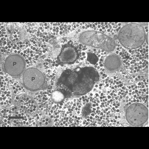
Normal morphology of two peroxisomes (marked P) in a human liver parenchymal cell. They are contiguous with a narrow endoplasmic reticulum cisterna; this relationship is frequent. The image also shows a large dark lysosome with a membrane, contrasted with cerium by acid phosphatase activity. Several mitochondria are seen, and in the cytosol many glycogen rosettes typical for liver of a fed person; and a few fat droplets (no membrane). Marker = 0.5 µm.

Technical Details

Prefixation in cold glutaraldehyde, acid phosphatase reaction with cerium, post fixation in OsO4.

Biological Sources
NCBI Organism Classification
Homo sapiens
Cell Type
parenchymal cell
Cellular Component
peroxisome
lysosome
mitochondrion
Biological Context
Biological Process
regulation of phosphatase activity
Attribution
Names
Frank Roels
Citation
Digital Object Identifier (DOI)
doi:10.7295/W9CIL46706
Archival Resource Key (ARK)
ark:/b7295/w9cil46706