Alternate header for print version


Licensing
Attribution Only: This image is licensed under a Creative Commons Attribution License. View License Deed | View Legal Code
tweet  
*CIL – Cell Image Library accession number. Please use this to reference an image.

CIL:46752*  Cite 
Description

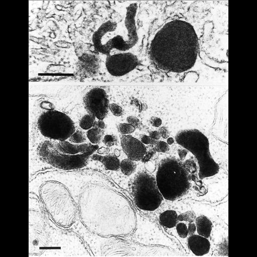
Peroxisomes in duodenal epithelial cells proliferate under treatment of oral phytol (a component of chlorophyll) over the course of 3 days. Phytol is oxidized to phytanic acid and pristanic acid, which may be the ultimate inducers. The cytosol contains free ribosomes. RER, SER and mitochondria are also seen. Bar = 0.5 µm.

Technical Details

Mice received phytol in their food (http://www.nature.com/pr/journal/v20/n5/full/pr198694a.html ). Peroxisomes are contrasted with DAB at pH 10.5 by their catalase activity, after fixation in buffered formal-calcium. Postfixation in Os04.

Biological Sources
NCBI Organism Classification
Mus musculus
Cell Type
epithelial cell
Cellular Component
peroxisome
Biological Context
Biological Process
peroxisome fission
Attribution
Names
Frank Roels
Published
"Peroxisomes. A personal account". pp. 151. Copyright VUB-Press 1991.
Citation
Digital Object Identifier (DOI)
doi:10.7295/W9CIL46752
Archival Resource Key (ARK)
ark:/b7295/w9cil46752