Alternate header for print version


Licensing
Attribution Non-Commercial Share Alike:This image is licensed under a Creative Commons Attribution, Non-Commercial Share Alike License. View License Deed | View Legal Code
tweet  
*CIL – Cell Image Library accession number. Please use this to reference an image.

CIL:47810*  Cite 
Description

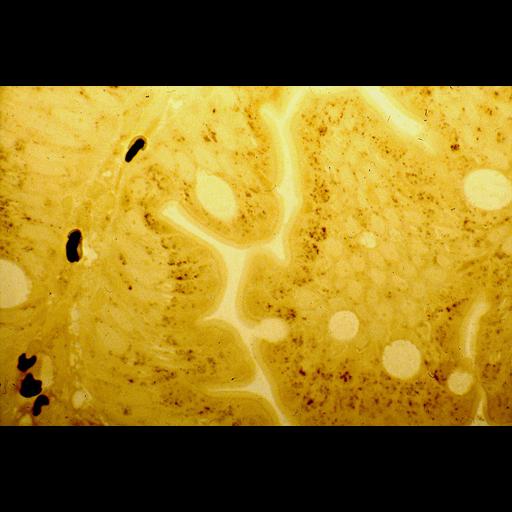
Human duodenal epithelium, at the level of the crypt. Fixation was carried out inbuffered formal-calcium. Peroxisomes are stained with DAB by their catalase activity at pH 10.5, postosmicated and embedded in epon. 2 µm plastic section.) Peroxisomes are the very small brown granules located between the brush border and the elongated epithelial nuclei (unstained). A few rounded goblet cells are recognized. In the subepithelial capillaries, 5 large black structures are erythrocytes that reacted with DAB by the peroxidatic activity of hemoglobin.

Technical Details

Bright field microscopy, immersion lens 100x

Biological Sources
NCBI Organism Classification
Homo sapiens
Cell Type
duct epithelial cell
goblet cell
erythrocyte
Cellular Component
peroxisome
Biological Context
Molecular Function
catalase activity
Attribution
Names
Frank Roels
Peter van der Spek
Marina Pauwels
Citation
Digital Object Identifier (DOI)
doi:10.7295/W9CIL47810
Archival Resource Key (ARK)
ark:/b7295/w9cil47810
Imaging
Image Mode
bright-field microscopy