Alternate header for print version


Licensing
Attribution Only: This image is licensed under a Creative Commons Attribution License. View License Deed | View Legal Code
tweet  
*CIL – Cell Image Library accession number. Please use this to reference an image.

CIL:48104*  Cite 
Description

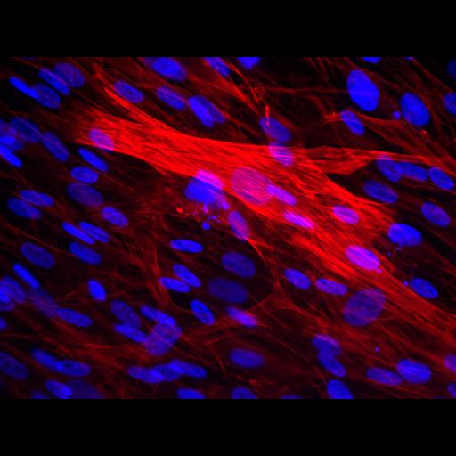
Cryopreserved rabbit skeletal muscle cells were revived and stained for actin (in red) to reveal cytoskeleton and DAPI (in blue) to label nuclei.

Technical Details

Rabbit skeletal muscle cells were fixed and immunostained for actin (from Santa Cruz), and DAPI (from Life Technologies). Images were acquired with a 40X (Plan Fluor, NA 0.75) lens, SPOT RT Slider CCD camera and SPOT Basic image capture software. Image generated with the Image J color combine function.

Biological Sources
NCBI Organism Classification
Rattus norvegicus
Cell Type
skeletal muscle cell
Cellular Component
actin filament
nucleus
Attribution
Names
Natalie Prigozhina
Citation
Digital Object Identifier (DOI)
doi:10.7295/W9CIL48104
Archival Resource Key (ARK)
ark:/b7295/w9cil48104
Imaging
Image Type
charge coupled device (CCD)
Image Mode
fluorescence microscopy
Parameters Imaged
fluorescence emission
Source of Contrast
distribution of epitope
Visualization Methods
4',6-diamidino-2-phenylindole (DAPI)
primary antibody plus labeled secondary antibody
Data Qualifiers
processed data
Sample Preparation
Methods
Dispersed cells cryopreserved
revived and cultured in vitro
Dimensions