Alternate header for print version


Licensing
Public Domain: This image is in the public domain and thus free of any copyright restrictions. However, as is the norm in scientific publishing and as a matter of courtesy, any user should credit the content provider for any public or private use of this image whenever possible. Learn more
tweet  
*CIL – Cell Image Library accession number. Please use this to reference an image.

CIL:7330*  Cite 
Description

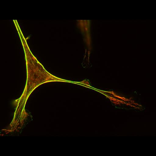
Primary aortic endothelial cells from transgenic mouse embedded in 3D collagen gels. Cell was fixed with 3% paraformaldehyde and stained with mouse antibody to phosphomyosin regulatory light chain (red), followed by anti-mouse Alexa 561 goat antibody and Alexa 488 phalloidin to stain for F-actin (green). Size and scale: 0.0789 micron/pixel, 1352x1040 pixels Images collected with a spinning disk confocal on a Nikon TE1000 with a 60X 1.2NA water objective using 300 ms exposure for the red channel and 200 ms for the green channel. See Fischer et al for details regarding preparation. http://www.ncbi.nlm.nih.gov/pubmed/19185493

Biological Sources
NCBI Organism Classification
Mus musculus
Cell Type
blood vessel endothelial cell
primary cell line cell
Cellular Component
actin cytoskeleton
myosin complex
Biological Context
Biological Process
actomyosin structure organization
Molecular Function
myosin phosphatase activity
Attribution
Names
Robert S. Fischer
Clare M. Waterman
Pubmed
19185493
OTHER
NHLBI/NIH
Citation
Digital Object Identifier (DOI)
doi:10.7295/W9CIL7330
Archival Resource Key (ARK)
ark:/b7295/w9cil7330
Sample Preparation
Methods
formaldehyde fixed tissue
Methods
dispersed cells in vitro
cells in 3D matrix
Dimensions
Spatial Axis Image Size Pixel Size
X 1352px 0.789µm
Y 1040px 0.789µm
Channel Wavelength
1 488,561nm