Alternate header for print version


Licensing
Public Domain: This image is in the public domain and thus free of any copyright restrictions. However, as is the norm in scientific publishing and as a matter of courtesy, any user should credit the content provider for any public or private use of this image whenever possible. Learn more
tweet  
*CIL – Cell Image Library accession number. Please use this to reference an image.

CIL:7428*  Cite 
Description

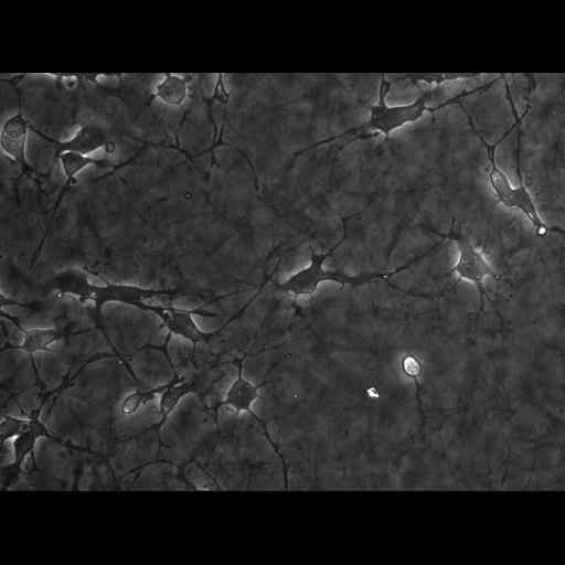
Primary endothelial cells from transgenic mouse embedded in 3D collagen gels of ~ 100 Pa Young's modulus are exposed to 10 micromolar blebbistatin (myosin II ATPase inhibitor). Phase contrast time lapse movie taken on a Nikon TE300 with a 20x 0.5 NA LWD objective. Images captured with an Orca II with 100 ms exposure. Image size is 0.1274 micron/pixel, 520x490 pixels, 20 seconds interval between timepoints.Time shown is in minutes:seconds See Fischer et al for details. http://www.ncbi.nlm.nih.gov/pubmed/19185493

Biological Sources
NCBI Organism Classification
Mus musculus
Cell Type
blood vessel endothelial cell
primary cell line cell
Cellular Component
cell
extracellular matrix
Biological Context
Biological Process
cell migration
Molecular Function
myosin II ATPase inhibition
Attribution
Names
Robert S. Fischer
Clare M. Waterman
Pubmed
19185493
OTHER
Laboratory of Cell and Tissue MorphodynamicsNHLBI / NIH
Citation
Digital Object Identifier (DOI)
doi:10.7295/W9CIL7428
Archival Resource Key (ARK)
ark:/b7295/w9cil7428
Imaging
Image Type
recorded image
Image Mode
phase contrast microscopy
Parameters Imaged
elastic scattering
Source of Contrast
differences in amount of elastic light scattering
Visualization Methods
visualization of contiguous regions
Processing History
unprocessed raw data
Sample Preparation
Methods
living tissue
Methods
dispersed cells in vitro
3D matrix
Dimensions
Spatial Axis Image Size Pixel Size
X 520px 0.1274——
Y 490px 0.1274nm