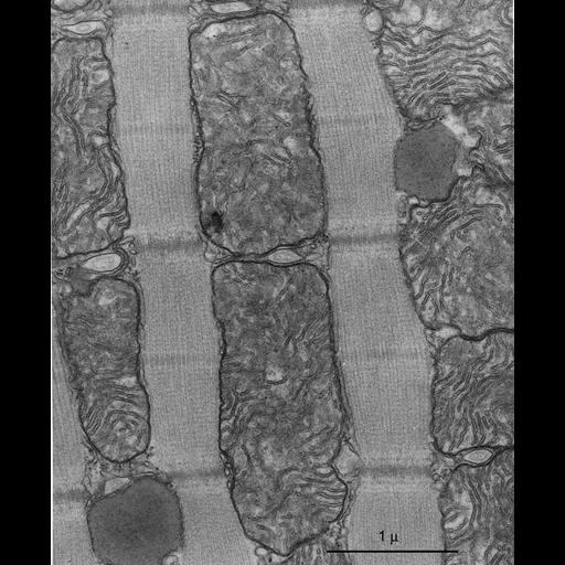
Cardiac muscle, particularly from the left ventricle, is rich in mitochondria since the heart requires an efficient continuous source of energy. In this highly magnified image, showing the length of two sarcomeres, large mitochondria occupy longitudinal spaces between the myofibrils. They are separated by transverse tubules and are associated with lipid droplets. Mitochondrial cristae, providing support of the enzymes of the respiratory cycle, are tightly packed in cardiac mitochondria. In the early 1960s, Keith Porter started collecting images, including this one, for an atlas of cell ultrastructure. This was one of the first such atlases to be published, and remains an extraordinary example of enduring quality. Interestingly, Porter selected to use tissues dissected from a bat for many of the images, because he knew that he could obtain excellent fixations. This image shows that his expectations were correct. Small strips from the working myocardium of the left ventricle of a bat were fixed by immersion in buffered glutaraldehyde, followed by buffered osmium and embedded in Epon. From: Porter KR, Bonneville M. Fine Structure of Cells and Tissues. 4th ed. Philadelphia, PA: Lea & Febiger; 1973. Original resource for this image, a 3 x 4 inch lantern slide, was provided by Keith R. Porter Archives (University of Maryland Baltimore County, Baltimore, MD). Digitization process: conversion from a 350 dpi tiff, 2729 x 3328 pixels. Research was conducted at Harvard University (Cambridge, MA). Original image was taken on February 28, 1963.