Alternate header for print version


Licensing
Public Domain: This image is in the public domain and thus free of any copyright restrictions. However, as is the norm in scientific publishing and as a matter of courtesy, any user should credit the content provider for any public or private use of this image whenever possible. Learn more
tweet  
*CIL – Cell Image Library accession number. Please use this to reference an image.

CIL:762*  Cite 
Description

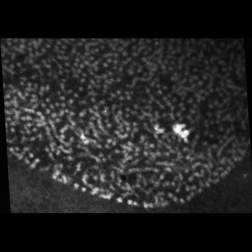
Upon electron bombardment, some elements, such as P, show an electron energy loss profile with a peak at a specific energy loss value. By collecting a series of images of a field at different energy losses, it is possible to compute the relative amount of the element at each pixel and map the element's distribution, a technique known as electron spectroscopic imaging (ESI) or electron filtered transmission EM (EFTEM). The 4 images in this group were recorded from unstained <30 nm section with 5 eV energy windows centered on 120, 104, 160 and 170 eV respectively using a Zeiss 902 energy filtered EM, making it possible to map the distribution of phosphorus (energy loss edge 160 eV) within the chromatin fibers. This image was recorded at 120 eV.

Biological Sources
NCBI Organism Classification
Patiria miniata
Cell Type
sperm
Cellular Component
chromatin
Biological Context
Biological Process
reproduction
Molecular Function
DNA compaction
Attribution
Names
Rachel Horowitz-Scherer
Chris Woodcock
Citation
Digital Object Identifier (DOI)
doi:10.7295/W9CIL762
Archival Resource Key (ARK)
ark:/b7295/w9cil762
Grouping This image is part of a group.
Imaging
Image Type
recorded image
Image Mode
microscopy with lenses
transmission electron microscopy (TEM)
Parameters Imaged
modes of electron microscopy
EELS imaging
Source of Contrast
electron scattering
Visualization Methods
electron energy loss window
Processing History
unprocessed raw data
Sample Preparation
Methods
glutaraldehyde fixed tissue
freeze substitution in Lowicryl
Relation To Intact Cell
sectioned tissue
Dimensions
Spatial Axis Image Size Pixel Size
X 4011px 0.4µm
Y 2804px 0.4µm