Alternate header for print version


Licensing
Public Domain: This image is in the public domain and thus free of any copyright restrictions. However, as is the norm in scientific publishing and as a matter of courtesy, any user should credit the content provider for any public or private use of this image whenever possible. Learn more
tweet  
*CIL – Cell Image Library accession number. Please use this to reference an image.

CIL:766*  Cite 
Description

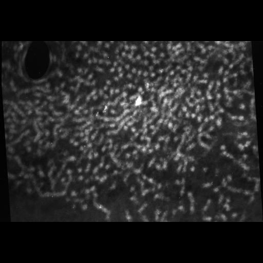
Phosphorus has a characteristic electron energy loss with an edge at 160 eV. The Zeiss 902 electron microscope permits images to be recorded at different energy losses which can be digitized and processed to yield a net phosphorus image. For nuclear chromatin, phosphorus is primarily located in DNA, allowing its distribution in chromatin fibers to be mapped. Electron energy loss (EELS) imaging is also known as electron spectroscopic imaging (ESI) and electron energy filtering TEM (EFTEM). Starfish sperm chromatin, which has clearly defined chromatin fibers was fixed, embedded in Lowicryl at low temperature, thin sections prepared and imaged unstained. The 4 grouped images were recorded with a Zeiss 902 energy filtered TEM at 80KV using 5eV windows centered on energy loss valuesof 170, 160, 120 (this image), and 104eV.

Biological Sources
NCBI Organism Classification
Patiria miniata
Cell Type
sperm
Cellular Component
nuclear chromatin
Biological Context
Biological Process
fertilization
Molecular Function
DNA packaging
Attribution
Names
Rachel Horowitz-Scherer
Chris Woodcock
Citation
Digital Object Identifier (DOI)
doi:10.7295/W9CIL766
Archival Resource Key (ARK)
ark:/b7295/w9cil766
Grouping This image is part of a group.
Imaging
Image Type
recorded image
Image Mode
transmission electron microscopy (TEM)
Parameters Imaged
modes of electron microscopy
EELS imaging
Source of Contrast
electron scattering
Visualization Methods
EELS window
Processing History
unprocessed raw data
Sample Preparation
Methods
glutaraldehyde fixed tissue
Low temperature embedding
Relation To Intact Cell
microtome-sectioned tissue
Dimensions
Spatial Axis Image Size Pixel Size
X 4072px 0.4nm
X 2804px 0.4nm