Alternate header for print version


Licensing
This image is copyright protected. Any public or private use of this image is subject to prevailing copyright laws. Please contact the content provider of this image for permission requests.
tweet  
*CIL – Cell Image Library accession number. Please use this to reference an image.

CIL:7722*  Cite 
Description

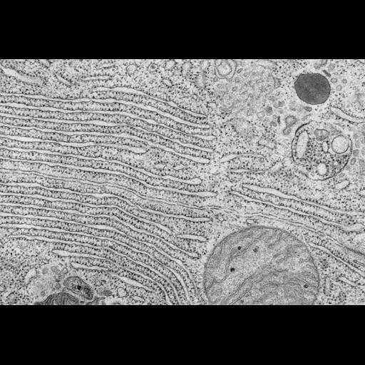
Stacks of rough endoplasmic reticulum (ER) cisternae seen in a transmission electron micrograph of a section through a secretory acinar cell from the bat exocrine pancreas. The rough ER is here involved in the synthesis of digestive enzymes that will be secreted by the pancreas and delivered to the duodenum, the initial segment of the small intestine. Each flattened ER cisterna has ribosomes associated with its cytoplasmic surface.

Technical Details

Small sections of bat pancreas were fixed by immersion in a primary buffered glutaraldehyde fixative, postfixed in buffered osmium tetroxide, dehydrated and embedded in Epon prior to examination in an electron microscope. Original resource provided by Keith R Porter Archives (University of Maryland Baltimore County, Baltimore, MD).Still image jp2; 12-May-67; 4x5 inch glass plate negative. This image was to be included as Figure 2-27 in the unpublished 5th edition of the Porter-Bonneville atlas: Fine Structure of Cells and Tissues.

Biological Sources
NCBI Organism Classification
Microchiroptera
Cell Type
acinar cell
pancreatic acinar cell
Cellular Component
rough endoplasmic reticulum
ribosome
Biological Context
Biological Process
digestion
secretion
Attribution
Names
Keith Porter
Mary Bonneville
OTHER
Keith R Porter Archives (University of Maryland Baltimore County, Baltimore, MD)
Citation
Digital Object Identifier (DOI)
doi:10.7295/W9CIL7722
Archival Resource Key (ARK)
ark:/b7295/w9cil7722
Imaging
Image Type
film
Image Mode
transmission electron microscopy (TEM)
detection of electrons
illumination by electrons
Parameters Imaged
electron density
Source of Contrast
differences in adsorption or binding of stain
Visualization Methods
osmium tetroxide
Processing History
digitized glass plate negative
Data Qualifiers
processed data
suitable for spatial measurements
Dimensions
Spatial Axis Image Size Pixel Size
X 4500px 21.1667µm
Y 3037px 21.1667µm