Alternate header for print version


Licensing
Public Domain: This image is in the public domain and thus free of any copyright restrictions. However, as is the norm in scientific publishing and as a matter of courtesy, any user should credit the content provider for any public or private use of this image whenever possible. Learn more
tweet  
*CIL – Cell Image Library accession number. Please use this to reference an image.

CIL:7744*  Cite 
Description

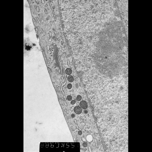
Details of a cryofixed Ptk tissue culture cell showing Golgi apparatus, mitochondria and a segment of nucleus with nucleolus at right. 40 nm epon section prepared from plunge frozen,and freeze substituted specimen. Image recorded at 55,000x with a Philips CM10 TEM operated at 80KV.

Biological Sources
NCBI Organism Classification
Potorous tridactylus tridactylus
Cell Type
primary cell line cell
Cell Line
Ptk1 kidney epithelium
Cellular Component
Golgi apparatus
nucleus
Attribution
Names
Mary Morphew
J. Richard McIntosh
Mark Ladinsky
Citation
Digital Object Identifier (DOI)
doi:10.7295/W9CIL7744
Archival Resource Key (ARK)
ark:/b7295/w9cil7744
Grouping This image is part of a group.
Imaging
Image Type
transmission electron microscopy (TEM)
film
Image Mode
illumination by electrons
detection of electrons
Parameters Imaged
electron density
Source of Contrast
electron-dense stain
Visualization Methods
uranyl salt
Processing History
unprocessed raw data
Sample Preparation
Methods
cryofixed tissue
freeze substitution
tissue in plastic embedment
Relation To Intact Cell
microtome-sectioned tissue
Dimensions
Spatial Axis Image Size Pixel Size
X 4268px 0.27nm
Y 6192px 0.27nm