Alternate header for print version


Licensing
Public Domain: This image is in the public domain and thus free of any copyright restrictions. However, as is the norm in scientific publishing and as a matter of courtesy, any user should credit the content provider for any public or private use of this image whenever possible. Learn more
tweet  
*CIL – Cell Image Library accession number. Please use this to reference an image.

CIL:9070*  Cite 
Description

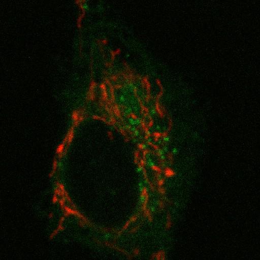
NRK cells expressing a mitochondria marker, mito-RFP (red), and PSS1-CFP (green), phosphatidylserine synthase 1. A single 6 micron confocal slice was imaged on a Zeiss LSM 510 every 5.9 sec with the follow parameters: Green: Ex: 458 nm laser, Em: Long Pass 475nm, Red: 543 nm laser, Em: Long pass 560 nm).

Biological Sources
NCBI Organism Classification
Rattus
Cell Type
fibroblast
Cell Line
NRK
Kidney
Cellular Component
mitochondrion
phosphatidylserine synthase 1
Biological Context
Biological Process
mitochondrion organization
Attribution
Names
Kasturi Mitra
Citation
Digital Object Identifier (DOI)
doi:10.7295/W9CIL9070
Archival Resource Key (ARK)
ark:/b7295/w9cil9070
Grouping This image is part of a group.
Imaging
Image Type
recorded image
Image Mode
single-spot confocal microscopy
Parameters Imaged
fluorescence emission
Source of Contrast
distribution of a specific protein
Visualization Methods
mRFP1
ECFP
Processing History
unprocessed raw data
color combine
Data Qualifiers
raw, unprocessed data
suitable for spatial measurements
Sample Preparation
Methods
living tissue
Relation To Intact Cell
dispersed cells in vitro
Dimensions
Spatial Axis Image Size Pixel Size
X 512px 0.07µm
Y 512px 0.07µm
Channel Wavelength
1 488, 543nm
Time 5.9 seconds