Alternate header for print version
Advanced search
Contributors
Help
Submit
Search
menu
Cell Process
Cell Component
Cell Type
Organism
Microbial
Alzheimer's
Data Sets
University of California, San Diego
9500 Gilman Drive
La Jolla, CA 92093-0608, USA
Voice
: (858) 534-0276
Fax
: (858) 534-7497
Email
: dorloff@ncmir.ucsd.edu
Reconstruction
Image Data Download Options...
Download in JPEG format
Download full resolution image
Download animation file
Open Detailed Viewer
Display image description
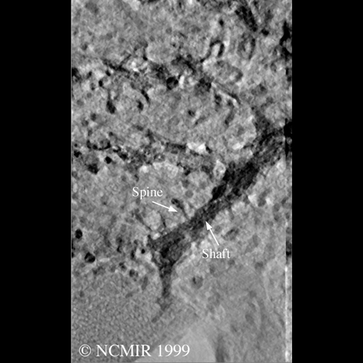
SIngle computed slice through tomographic volume of selectively stained Purkinje cell spiny dendrite from rat cerebellar cortex.
Full resolution image description
Zip file containing compressed volume in Analyze 7.5 format. File contains both .hdr and .img files.
Volume_dimension
390, 640, 220
Volume scale
0.021, 0.021, 0.021
Animation description
Animation through computed slices of tomographic reconstruction of a selectively stained Purkinje cell spiny dendrite.
Segmentation
Image Data Download Options...
Download in JPEG format
Download segmentation file
Segmentation file description
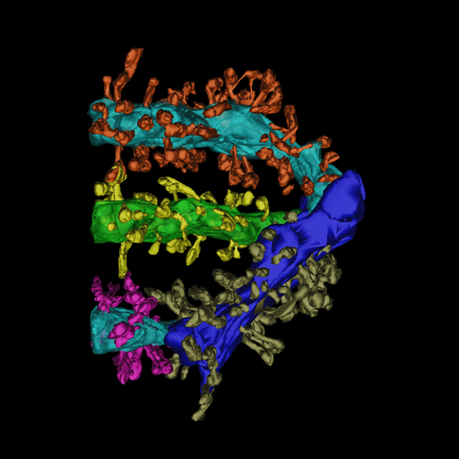
Zip file containing the original trace file (osaka1.r.trace), the surfaced objects (*.synu) and the Viewdata file required to view them using Synu.
License
Attribution Only:
This image is licensed under a Creative Commons Attribution License.
View License Deed
|
View Legal Code
CCDB:3494
*
Cite
Project:
P1119
Project name
Correlated Microscopy of Dendritic Spines
Description
Measurements of spine parameters using light microscopy and electron tomography
Funding agency
NIH
Leader(s)
Maryann Martone
Collaborator(s)
Naoko Yamada; Gordun Arbuthnott; Cali Ingham; Stephen Young
Start date
01-01-1992
End date
unspecified
Experiment
Experiment ID
10
Title
spiny dendrite
Purpose
how well dendritic spines can be detected and measured using LM
Experimenter(s)
Naoko Yamada
Microscopy product
Microscopy product ID
3494
Instrument
Hitachi 3MeV UHVEM
Microscopy type
UHVEM
Product type
SINGLE TILT
Image basename
osaka1r
Subject
Species
rat
Scientific name
rattus norvegicus
Strain
Sprague Dawley
Treatment
none
Age class
adult
Tissue section
Anatomical location
cerebellum
Microtome
Ultramicrotome
Thickness
4 µm
Specimen description
Organ
brain
System
central nervous system
Structure
spiny dendrite
Cell type
Purkinje neuron
Imaging parameters
Type
Electron microscopy product
Recording medium
film
Magification
4000
Accelerating voltage
3 MeV
Specimen preparation
Protocol used
Intracellular injection with Lucifer Yellow followed by photooxidation.
Imaging product type
Type
Single tilt
Description
singlet_desc
Min range
-60 degrees
Max range
60 degrees
Notes
Specimen was pre-irradiated prior to imaging
×
Citation Information
Maryann Martone, Naoko Yamada; Gordun Arbuthnott; Cali Ingham; Stephen Young (1992) CCDB:3494, rattus norvegicus, spiny dendrite, Purkinje neuron. CIL. Dataset. https://doi.org/doi:10.7295/W9CCDB3494