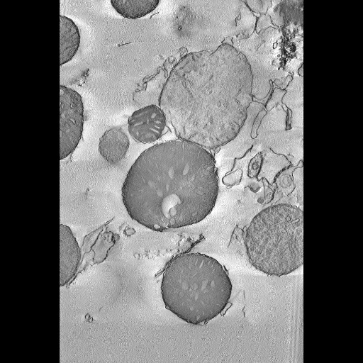
Computed slice through a tomographic volume of isolated mitochondria from mouse liver after treatment with the BH3 peptide domain from Bid.
Full resolution image description
Zip file (rudyE_vol.zip) containing the reconstruction file in Analyze 7.5 format (RudyEsmall.img/hdr). Note: This volume was likely downsampled from the original volume and thus the stated voxel size is likely incorrect. The full sized volume file could not be located. Also note, a subvolume was generated from this reconstruction for segmentation (RudyE_scirun.img/hdr). The subvolume used for tracing may be downloaded along with the other segmentation files. The location of the subvolume can be viewed in the image map stored with this record.
Volume_dimension
673, 1002, 321
Volume scale
0.0014, 0.0014, 0.0014
Animation description
Animation through a portion of the tomographic volume of isolated mitochondria treated with the BH3 peptide domain from Bid. The volume was downsampled for ease of viewing.
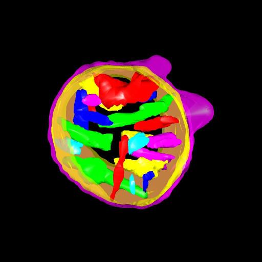
Manual segmentation of the individual cristae and inner and outer mitochondrial membranes using Xvoxtrace 2.18. Contours were traced every fourth section starting on plane 4
Segmentation file description
Zip file (RudyE_seg.zip) containing the segmentation file generated by Xvoxtrace and converted into Jinx format (RudyE_seg.jnx), the subvolume used for tracing in Analyze 7.5 format (RudyE_scirun.img/.hdr) and the surfaced objects in Synu format (*.synu). Note: For unknown reasons, the .trace file originally generated by Xvoxtrace could not be uploaded. The Analyze 7.5 volumes were also acting a bit strange, but could be uploaded after regenerating them using ImageJ. Both the segmentation and volume files could be opened in Jinx, and so the manual segmentations were saved as a Jinx file. Jinx can be downloaded from the CCDB tool web page.
Proapoptotic BH3-only proteins induce Bax/Bak-dependent mitochondrial cristae remodeling independent of cytochrome c release and Bak oligomerization
Leader(s)
Ryuji Yamaguchi
Ph.D.
Collaborator(s)
Don Newmeyer; Lydia Lartigue; Guy Perkins
Ph.D.; Ray T Scott; Amruta Dixit;
Start date
unspecified
End date
unspecified
Experiment
Experiment ID
3458
Title
Cytochrome C Release Assay
Purpose
To determine the effect of pro-apoptotic peptides on mitochondrial morphology and cytochrome C release.
Experimenter(s)
Guy Perkins
Microscopy product
Microscopy product ID
4097
Instrument
JEOL4000EX IVEM
Microscopy type
IVEM
Product type
SINGLE TILT
Image basename
Rudy E
Subject
Species
mouse
Scientific name
mus musculus
Strain
unknown
Group by
Drug treatment
Treatment
Mitochondria were incubated with Bid derived BH3 peptides at 37 degrees C for 12 min in AT buffer containing 10 mM KCl and centrifuged at 5200x g for 6 min.
Age class
adult
Tissue section
Anatomical location
liver
Tissue product storage
liquid nitrogen
Thickness
0.5 µm
Specimen description
Organ
liver
Structure
mitochondrion
Imaging parameters
Type
Electron microscopy product
Recording medium
film
Magification
20000
Accelerating voltage
400 KeV
Specimen preparation
Protocol used
Mouse liver mitochondria were prepared as described (Yamaguchi et al., 2006).Pelleted mitochondria were fixed with a 37 C solution of 2% paraformaldehyde, 2.5% glutaraldehyde (Ted Pella) in 0.15 M sodium cacodylate (pH 7.4), and then incubated for an additional 30 minutes on ice. Fixed samples were then rinsed 3 times for 3 minutes each with 0.15 M sodium cacodylate plus 3 mM calcium chloride (pH 7.4) on ice, post-fixed with 1 percent osmium tetroxide, 0.8% potassium ferrocyanide, 3 mM calcium chloride in 0.15 M sodium cacodylate (pH 7.4) for 60 minutes, and then washed 3 times for 3 minutes with ice-cold distilled water. The samples were stained overnight with 2% uranyl acetate at 4 degrees C, dehydrated in graded ethanol baths, and embedded in Durcupan ACM resin (Fluka).Sections from the embedded mitochondria samples were cut at thicknesses of nominally 500 nm. Sections were then stained 30 min in 2 percent aqueous uranyl acetate, followed by 15 min in lead salts. Fiducial cues consisting of 15 and 20 nm colloidal gold particles were deposited on opposite sides of the section
Imaging product type
Type
Single tilt
Description
singlet_desc
Min range
-60 degrees
Max range
60 degrees
Tilt increment
2 degrees
Notes
tilted using a computer controlled goniometer about an axis perpendicular to
the optical axis of the microscope
Citation Information
Ryuji Yamaguchi, Ph.D., Don Newmeyer; Lydia Lartigue; Guy Perkins, Ph.D.; Ray T Scott; Amruta Dixit; CCDB:4097, mus musculus, mitochondrion. CIL. Dataset. https://doi.org/doi:10.7295/W9CCDB4097