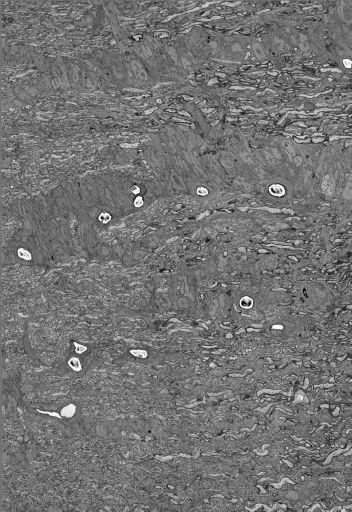
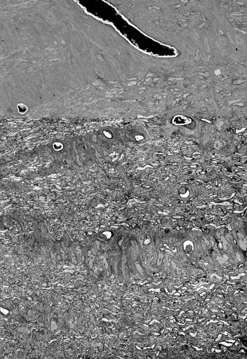
Single slice through a serial section reconstruction of the optic nerve head in the area of transition between myelinated and unmyelinated axons the mouse obtained through serial section scanning electron microscopy. The location of the transition zone may be viewed on the image map (red box) associated with this data set.
Full resolution image description
Stitched, normalized reconstruction in IMOD format. Data set was normalized by computing the mean and standard deviation of the entire dataset then setting each plane to that mean and standard deviation by subtracting from each voxel the old mean (that is, the original mean of the plane), dividing by the old standard deviation, then multiplying by the new standard deviation and adding in the new mean. The location of the transition zone may be viewed on the image map (red box) associated with this data set.
**Note: due to the size of the file (>200Gb), this data set is not available for automated download from the CCDB. A downsampled version (9Gb) may be downloaded. The full resolution data may be viewed through the WIB tool. If you would like the full resolution data file, please contact CCDB.
Downsampled image description
Binned version of the normalized volume in mrc format.
pixel dwell time = 8 usec
chamber pressure = High Vacuum
spot size = 3.0
Specimen preparation
Protocol used
The animal was perfused with Ringer's followed by 2% PFA / 2.5% glutaraldehyde. The tissue was postfixed in the same fixative for 2 hours on ice. The eye was enucleated and the optic nerve head was dissected for sagittal longitudinal section.The tissue was placed into solution containing 3% potassium dichromate and 2% osmium tetroxide in ddH2O. The samples were allowed to sit at room temp for 3 hours. The tissue washed with ddH2O at room temp 3x 5 minutes and placed in filtered thiocarbohydrazide(TCH) solution(0.1 g TCH in 10 mL ddH2O) for 30 minutes at room temp.The tissue washed with ddH2O at room temp 3x 5 minutes and placed in 2% osmium in ddH20 for an hour at room temp.The tissue washed with ddH2O at room temp 3x 5 minutes and placed in 1% aq. UA overnight in fridge.The next day, dissolve 0.066g of lead nitrate in 10 mL aspartic acid solution and adjust pH to 5.5 with 1N KOH. Place solution in 60 degree oven for 30 minutes. No precipitate should form.Take tissue from 1% UA and wash with ddH2O, 3x 5 minutes.Place tissue in the lead aspartate solution and leave in oven for 30 minutes. Wash tissue 3x 5 minutes with ddH2O and dehydrate and embed in Durcupan as described below.The tissue was dehydrated in EtOH (20, 50, 70, 90, 100, 100% for 10 minutes each) and then acetone (100% 3x 10 minutes). The tissue was placed into 50:50 Durcupan:acetone overnight. Tissue was placed in 100% Durcupan for 1 hour. Tissue was transferred to fresh Durcupan for 2 hours. Tissue was placed into 60 degree oven for 2 days and polymerized.
Imaging product type
Type
Mosaic
Description
mosaic with 6 sections
X position
2 tiles
Y position
3 tiles
Imaging product type
Type
Serial section
Cutting plane
longitudinal
Z resolution
70 nm/pixels
Notes
serial section series and mosaic
Citation Information
Keun-Young Kim, Eric Bushong, Mark Ellisman, Daniela Boassa, Masako Terada, Nicholas Marsh-Armstrong (2010) CCDB:8093, Mus musculus. CIL. Dataset. https://doi.org/doi:10.7295/W9CCDB8093