Xvoxtrace file containing manually contours produced from volume available under the reconstruction section. No surface renderings were found associated with this file.
In situ structures of mitochondria in rods and cones
Leader(s)
Don Fox
University of Houston
Collaborator(s)
Guy Perkins
Start date
09-01-2001
End date
09-01-2001
Experiment
Experiment ID
33
Experiment date
07-01-2001
Title
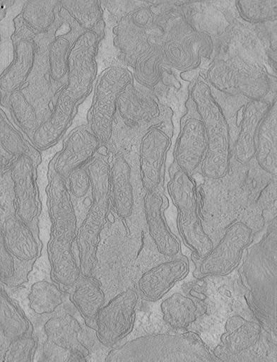
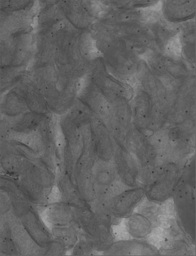
Cone and rod mitochondria: electron tomography
Purpose
electron tomography of cone mitochondria
Experimenter(s)
Guy Perkins
Microscopy product
Microscopy product ID
8752
Instrument
JEOL4000EX
Microscopy type
IVEM
Product type
SINGLE TILT
Image basename
OD
Subject
Species
mouse
Scientific name
mus musculus
Strain
C57BL/6
Treatment
none
Age class
adult
Tissue section
Anatomical location
retina
Specimen description
Organ
eye
System
central nervous system
Structure
mitochondrion
Cell type
retina rod cell
Imaging parameters
Type
Electron microscopy product
Magification
0
Accelerating voltage
400 KeV
Specimen preparation
Protocol used
These specimens were prepared similarly to those described in the Perkins et al. 2003 study (see project info), although the reconstruction did not appear in this paper.
Imaging product type
Type
Single tilt
Description
singlet_desc
Min range
-60 degrees
Max range
60 degrees
Tilt increment
2 degrees
Citation Information
Don Fox, University of Houston, Guy Perkins (2001) CCDB:8752, mus musculus, mitochondrion, retina rod cell. CIL. Dataset. https://doi.org/doi:10.7295/W9CCDB8752