Alternate header for print version
Advanced search
Contributors
Help
Submit
Search
menu
Cell Process
Cell Component
Cell Type
Organism
Microbial
Alzheimer's
Data Sets
Center for Research in Biological Systems
University of California, San Diego
9500 Gilman Drive
La Jolla, CA 92093-0608, USA
Voice
: (858) 534-0276
Fax
: (858) 534-7497
Email
: dorloff@ncmir.ucsd.edu
Search Results for
microscopy
(11125 results)
(Not the results you were expecting?)
(Comments)
Still Images
Video/Animation
Z-Stack
Time Series
CIL:36022
NCBI Organism Classification
Chlorocebus aethiops
Biological Process
negative regulation of insulin receptor signaling pathway
Cellular Component
none specified
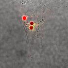
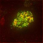
COS7 cells, transfected with GFP-PTP1B and APN-EphA3, when co-transfected with TM-BirA, yielded biotinylated EphA3 that can be stimulated with SA-Dynabeads. GFP-PTP1B and EphA3-transfected COS7 cells...
CIL:13654
NCBI Organism Classification
Homo sapiens
Biological Process
Golgi organization
Cellular Component
trans-Golgi network
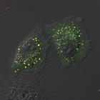
Image is Fig. 4D in PMID: 20679433. HeLa cells were transfected to co-express p125A-mCherry and Sec31A-GFP. p125A colocalizes extensively with Sec31A; live cell time-lapse video microscopy showed th...
CIL:50453
NCBI Organism Classification
Mus musculus
Biological Process
none specified
Cellular Component
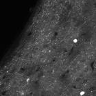
Mouse brain tissue expressing tdTomato in corticotropin releasing factor-expressing neurons
In this study, we developed an electron microscopy method, called CryoChem, to optimally preserve the morphology of genetically labeled tissues. This method also allows us to perform 3D correlated lig...
CIL:50454
NCBI Organism Classification
Mus musculus
Biological Process
none specified
Cellular Component
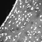
DNA in the nucleus is stained with DRAQ5
In this study, we developed an electron microscopy method, called CryoChem, to optimally preserve the morphology of genetically labeled tissues. This method also allows us to perform 3D correlated lig...
CIL:35463
NCBI Organism Classification
Rattus norvegicus
Biological Process
none specified
Cellular Component
none specified
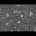
Phase contrast microscopy of liver sinusoidal endothelial cells purified from a Brown Rat.
CIL:38680
NCBI Organism Classification
Barbulanympha
Biological Process
mitosis
Cellular Component
spindle
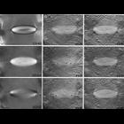
The series of three vertical columns shows various aspects of mitotic anaphase in a live Barbulanympha (protist). The left column shows the birefringent spindle imaged by polarization microscopy; the...
CIL:23049
NCBI Organism Classification
Rattus
Biological Process
gelsolin treatment
Cellular Component
microtubule
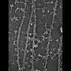
Association of plectin with MTs. Electron microscopy of gelsolin-treated REF-52 cells after immunogold (10 nm) labeling for plectin. Plectin forms bridges between two microtubules. Electron microscopy...
CIL:12594
NCBI Organism Classification
Homo sapiens
Biological Process
none specified
Cellular Component
cell
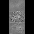
A human (Homo sapien) cheek cell smeared on a coverslip and imaged by birghtfield, phase contrast and differential interference contrast (DIC) microscopy. Images were acquired on a Zeiss Axiovert 200M...
CIL:40753
NCBI Organism Classification
Canis lupus familiaris
Biological Process
protein targeting
Cellular Component
P-75
Total internal reflection (TIRF) micrograph of MDCK cells stably transfected with the apical membrane protein P75-GFP (green) and with its sorting receptor Galectin-3-dsRed (red) in close proximity or...
CIL:50401
NCBI Organism Classification
Mus musculus
Biological Process
none specified
Cellular Component
none specified
In this study, we developed an electron microscopy method, called CryoChem, to optimally preserve the morphology of genetically labeled tissues. This method also allows us to perform 3D correlated lig...
1
2
3
4
5
6
7
8
9
...
1113
Next »
Results per page:
10
20
50
100