Alternate header for print version
Advanced search
Contributors
Help
Submit
Search
menu
Cell Process
Cell Component
Cell Type
Organism
Microbial
Alzheimer's
Data Sets
Center for Research in Biological Systems
University of California, San Diego
9500 Gilman Drive
La Jolla, CA 92093-0608, USA
Voice
: (858) 534-0276
Fax
: (858) 534-7497
Email
: dorloff@ncmir.ucsd.edu
Search Results for
Nerve cell layer
(59 results)
(Not the results you were expecting?)
(Comments)
Still Images
Video/Animation
Z-Stack
Time Series
CIL:39022
NCBI Organism Classification
Danio rerio
Biological Process
optic nerve structural organization
Cellular Component
neuron projection
Confocal micrograph showing the connections of the visual system in a four-day-old zebrafish embryo. Staining of the neurons, glia and optic nerve illustrate the connections between the retina and the...
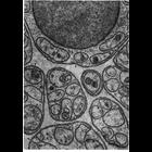
CIL:10950
NCBI Organism Classification
Felis catus
Biological Process
plasma membrane organization
Cellular Component
basement membrane
Schwann cells in peripheral nerves secrete a layer similar to the basal lamina called the lamina externa, boundary layer, or basement membrane. In this micrograph, the lamina externa surrounds the Sc...
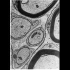
CIL:10948
NCBI Organism Classification
Rattus
Biological Process
cell-substrate adhesion
Cellular Component
basement membrane
Electron micrograph of a nerve from the mesentery of a rat shows groups of unmyelinated axons wrapped by deeply invaginating Schwann cells in cross section. Surrounding the Schwann cell wrapping is a ...
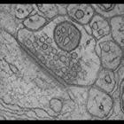
CIL:40020
NCBI Organism Classification
Rattus norvegicus
Biological Process
none specified
Cellular Component
synapse
Single computed slice through a tomographic reconstruction of a spine synapse from the molecular layer of the cerebellar cortex from tissue that was prepared from a combination of chemical fixation an...
CIL:10113
NCBI Organism Classification
Rattus
Biological Process
developmental process
Cellular Component
cytoskeleton
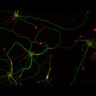
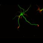
This multi-layer image shows the spatial relationship between filamentous actin (red) and microtubule array (green) in cultured hippocampal neurons, grown for 5 days in vitro. Actin staining (with rh...
CIL:10208
NCBI Organism Classification
Rattus
Biological Process
developmental process
Cellular Component
cytoskeleton
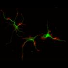
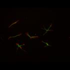
This multi-layer image shows the spatial relationship between filamentous actin (red) and microtubule array (green) in cultured hippocampal neurons, grown for 1 day in vitro. Actin staining (with rho...
CIL:10211
NCBI Organism Classification
Rattus
Biological Process
developmental process
Cellular Component
cytoskeleton
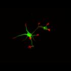
This multi-layer image shows the spatial relationship between filamentous actin (red) and microtubule array (green) in cultured hippocampal neurons, grown for 1 day in vitro. Actin staining (with rho...
CIL:10218
NCBI Organism Classification
Rattus
Biological Process
developmental process
Cellular Component
cytoskeleton
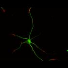
This multi-layer image shows the spatial relationship between filamentous actin (red) and microtubule array (green) in cultured hippocampal neurons, grown for 3 days in vitro. Actin staining (with rh...
CIL:10224
NCBI Organism Classification
Rattus
Biological Process
developmental process
Cellular Component
cytoskeleton
This multi-layer image shows the spatial relationship between filamentous actin (red) and microtubule array (green) in cultured hippocampal neurons, grown for 3 days in vitro. Actin staining (with rh...
CIL:10095
NCBI Organism Classification
Rattus
Biological Process
developmental process
Cellular Component
cytoskeleton
This multi-layer image shows the spatial relationship between filamentous actin (red) and microtubule array (green) in cultured hippocampal neurons, grown for 1 day in vitro. Actin staining (with rho...
1
2
3
4
5
6
Next »
Results per page:
10
20
50
100