Alternate header for print version
Advanced search
Contributors
Help
Submit
Search
menu
Cell Process
Cell Component
Cell Type
Organism
Microbial
Alzheimer's
Data Sets
University of California, San Diego
9500 Gilman Drive
La Jolla, CA 92093-0608, USA
Voice
: (858) 534-0276
Fax
: (858) 534-7497
Email
: dorloff@ncmir.ucsd.edu
Reconstruction
Image Data Download Options...
Download in JPEG format
Download full resolution image
Open Detailed Viewer
Display image description
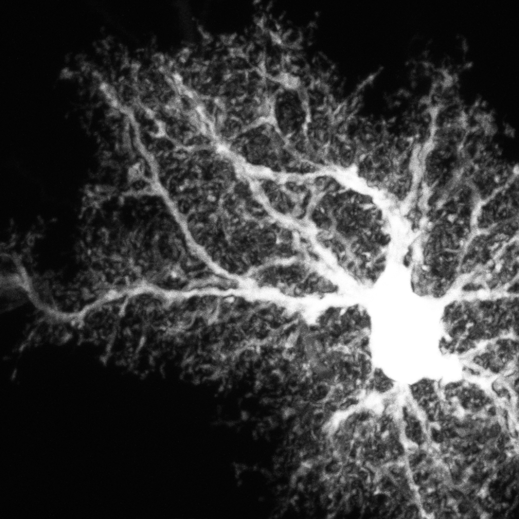
Optical section series through a protoplasmic astrocyte in rat hippocampal area CA1 intracellulaly injected Lucifer Yellow and imaged with confocal microscopy
License
Attribution Only:
This image is licensed under a Creative Commons Attribution License.
View License Deed
|
View Legal Code
CCDB:1028
*
Cite
Project:
P1230
Project name
Astrocyte Development
Description
Postnatal development of protoplasmic astrocytes
Funding agency
NIH
Leader(s)
Eric Bushong
Collaborator(s)
Maryann Martone
Mark Ellisman
Start date
02-01-2002
End date
unspecified
Experiment
Experiment ID
22
Title
Morphology of astrocytes in 3 week old hippocampus
Purpose
Examine the morphology of 3 week old astrocytes
Experimenter(s)
Eric Bushong
Microscopy product
Microscopy product ID
1028
Instrument
Biorad Radiance2000
Microscopy type
single photon confocal
Product type
optical section series
Image basename
3wk-ly23-hm
Spatial Axis
Image Size
Pixel Size
X
1024px
0.027407 µm
Y
1024px
0.027407 µm
Subject
Species
rat
Scientific name
rattus norvegicus
Strain
Sprague Dawley
Group by
NA
Treatment
NA
Age
3 weeks
Age class
juvenile
Tissue section
Anatomical location
hippocampus
Microtome
vibratome
Tissue product storage
coverslipped
Thickness
100 µm
Specimen description
Organ
brain
System
central nervous system
Cell type
protoplasmic astrocyte
Imaging parameters
Type
Light microscopy product
Immersion medium
oil
Mounting medium
gelvatol
Lens
Nikon
Lens magnification
X
Numerical aperture
1.4
Refractive index
1.5
Imaging product type
Type
Optical section
Z resolution
0.1 um
×
Citation Information
Eric Bushong, Maryann Martone, Mark Ellisman (2002) CCDB:1028, rattus norvegicus, protoplasmic astrocyte. CIL. Dataset. https://doi.org/doi:10.7295/W9CCDB1028