Alternate header for print version
Advanced search
Contributors
Help
Submit
Search
menu
Cell Process
Cell Component
Cell Type
Organism
Microbial
Alzheimer's
Data Sets
University of California, San Diego
9500 Gilman Drive
La Jolla, CA 92093-0608, USA
Voice
: (858) 534-0276
Fax
: (858) 534-7497
Email
: dorloff@ncmir.ucsd.edu
Reconstruction
Image Data Download Options...
Download in JPEG format
Download full resolution image
Download animation file
Open Detailed Viewer
Display image description
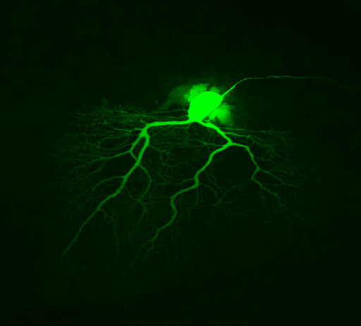
Purkinje neuron from mouse cerebellum injected with Lucifer Yellow and imaged using confocal microscopy
Volume_dimension
1024, 1024, 89
Animation description
Rotation loop of a maximum intensity projection of a Purkinje neuron injected with Lucifer Yellow, rotated along the y axis
License
Attribution Only:
This image is licensed under a Creative Commons Attribution License.
View License Deed
|
View Legal Code
CCDB:20
*
Cite
Project:
P1173
Project name
CCDB rat test data pt. 2
Description
Confocal images
Funding agency
NIH
Leader(s)
Maryann Martone
Collaborator(s)
Diana Price and Andrea
Start date
05-15-2002
End date
unspecified
Experiment
Experiment ID
12
Experiment date
05-15-2002
Title
Intracellular injection of Purkinje neuron
Purpose
To obtain multi resolution data for CCDB
Experimenter(s)
Diana Price and Andrea Thor and M. Terada
Microscopy product
Microscopy product ID
20
Instrument
Biorad Radiance 2000 Confocal
Microscopy type
confocal
Product type
optical section series
Image basename
e1cb10a3
Spatial Axis
Image Size
Pixel Size
X
1024px
Y
1024px
Subject
Species
rat
Scientific name
rattus norvegicus
Strain
Sprague Dawley
Treatment
none
Age
1 months
Age class
adult
Tissue section
Anatomical location
cerebellum
Microtome
vibratome
Thickness
100 µm
Specimen description
Organ
brain
System
central nervous system
Map location
View location
Cell type
Purkinje neuron
Atlas coordinate
0, 0, 0,
Imaging parameters
Type
Light microscopy product
Immersion medium
oil
Mounting medium
gelvatol
Lens magnification
X
Numerical aperture
1.3
Imaging product type
Type
Optical section
Z resolution
0.25 um
×
Citation Information
Maryann Martone, Diana Price and Andrea (2002) CCDB:20, rattus norvegicus, Purkinje neuron. CIL. Dataset. https://doi.org/doi:10.7295/W9CCDB20