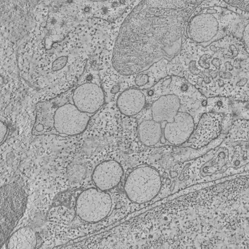
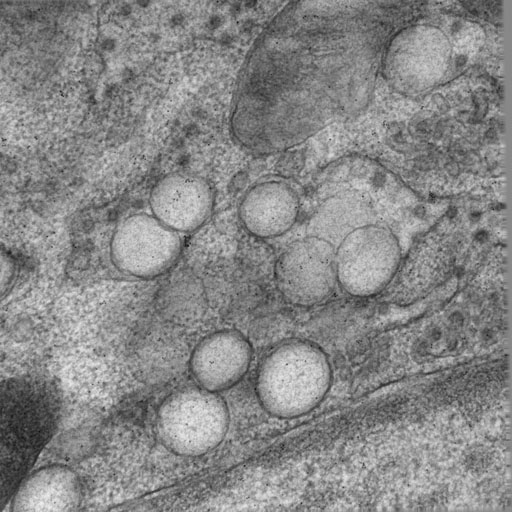
Alternate header for print version


Reconstruction
Display image description
Single computed slice through a dual axis tilt electron tomographic reconstruction of a freeze-substituted VeroE6 cultured cell infected with the SARS-CoV.
Full resolution image description
Tomographic volume in IMOD format (6022_Suppmovie3.rec) of the infected VeroE6 cells.
Volume_dimension
2023, 2023, 167
Volume scale
0.0012, 0.0012, 0.0012
Animation description
Animation (6022.avi) through the tomographic volume of freeze-substituted VeroE6 culture cells infected with SARS-CoV.

Image 2D
Display image description
Single tilt image from a double tilt series through 0.2 um thick section of a freeze-substituted VeroE6 cultured cell infected with the SARS-CoV taken with transmission electron microscopy. Dark particles are 10 nm colloidal gold particles that were applied to the surface to serve as fiducial markers for alignment of the tilt series.
Full resolution image description
Aligned tilt images (6022_Suppmovie3.ali) in IMOD format

License
Attribution Only: This image is licensed under a Creative Commons Attribution License. View License Deed | View Legal Code

CCDB:6022*  Cite 
Project: P2005
Project name
Membrane Modifications Induced by Corona Virus
Description
Electron tomography of African Green Monkey epithelial cells infected with SARS-coronavirus
Funding agency
Leiden University Medical Center (LUMC, the Netherlands), Council for Chemical Sciences of the Netherlands Organization for Scientific Research (NOW-CW grants 700.52.306 and 700.55.002), European Commission under context of Euro-Asian SARS-DTV Network (SP22-CT-2004-511064)
Leader(s)
Kèvin Knoops
Abraham J. Koster
A. Mieke Mommaas and Eric J. Snijder
Collaborator(s)
Marjolein Kikkert
Sjoerd H.E. van den Worm
Jessika C. Zevenhoven-Dobbe
Yvonne van der Meer
Start date
02-01-2005
End date
02-01-2005
 
Experiment
Experiment ID
6014
Experiment date
06-22-2006
Title
Electron Tomography of Membrane Modification
Purpose
Study the membrane modification of African Green Monkey epithelial cells post-infection of SARS-coronavirus
Experimenter(s)
Kevin Knoops
Microscopy product
Microscopy product ID
6022
Instrument
FEI Tecnai 12
Microscopy type
TEM
Product type
DOUBLE TILT
Image basename
suppmovie3
Spatial Axis Image Size Pixel Size
X 2023px 1.2 nm/pixels
Y 2023px 1.2 nm/pixels
Subject
Species
monkey
Scientific name
Cercopithecus aethiops
Strain
Vervet
Group by
time after infection
Treatment
7 hrs post-infection with SARS-CoV strain Frankfurt-1 (provided by Dr. H. F. Rabenau 603 and Dr. H. W. Doerr (Johann-Wolfgang-Goethe-Universität, Frankfurt am Main, Germany)
Age class
n/a
Tissue section
Anatomical location
epithelium
Microtome
Leica UC6 ultramicrotome
Thickness
0.2 µm
Specimen description
Organ
kidney
Cell type
VeroE6
Imaging parameters
Type
Electron microscopy product
Recording medium
Slow scan cooled 2K CCD camera
Magification
18500
Accelerating voltage
120 kV
Specimen preparation
Protocol used
For ultrastructural morphological investigations, SARS-CoV-infected Vero E6 cells were pre-fixed (for biosafety reasons) overnight with 3% paraformaldehyde in 0.1M PHEM buffer (60 mM piperazide-1,4-bis[2-ethanesulfonic acid], 25 mM HEPES, 2 mM MgCl2, 10 mM EGTA) at various time points after infection. For cryo-fixation, cell monolayers adhered to Thermanox coverslips (Nunc, Denmark) were plunged into liquid ethane. Freeze substitution was performed at -90C in an automated freeze-substitution system (Leica, Austria) using an FS medium consisting of 90% acetone and 10% water, containing 1% osmium tetroxide and 0.5 % uranyl acetate. After washing with pure acetone at RT, the samples were embedded in epoxy LX-12 resin. Thin sections were collected on 100 Mesh grids, 1.25% pioloform support film contrasted with uranyl acetate and lead hydroxide and subsequently viewed at 80 kV with a Philips CM-10 transmission electron microscope (Philips, The Netherlands).
Imaging product type
Type
Double tilt
X min range
-65 degrees
X max range
65 degrees
X tilt increment
1 degrees
Y min range
-65 degrees
Y max range
65 degrees
Y tilt increment
1 degrees
Description
specimens were rotated 90 deg around the Z-axis using a dual-axis tilt tomography holder (Fishione L