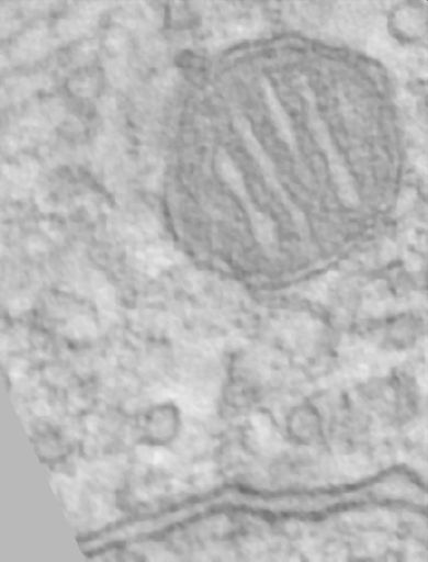
Single computed slice from a tomographic reconstruction through the Calyx of Held of an adult cat. The mitochondrion in the presynaptic terminal exhibits a structural specialization termed the mitochondrial adherens complex.
Full resolution image description
Tar file containing the volume in Analyze 7.5 format. Both the .img (queen_sub.img_ and .hdr (queen_sub.hdr) files are included.
File format
MRC
Volume_dimension
435, 570, 250
Animation description
Animation through the computed slices of a tomographic reconstruction through the Calyx of Held of an adult cat. The mitochondrion in the presynaptic terminal exhibits a structural specialization termed the mitochondrial adherens complex.
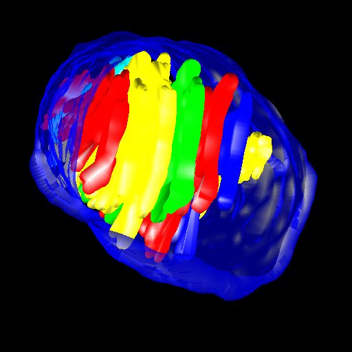
Manual segmentation of mitochondrial membranes using Xvoxtrace followed by surfacing using Synu
Segmentation file description
Segmentation files including the manual contours generated with Xvotrace (.trace) and and surfaced objects in Synu format (.synu, Viewdata), and .surf files. Note: the volume referenced in the Xvoxtrace file was no longer available. The reconstruction provided in the Reconstruction record should work.
Electron tomography of mitochondrial adherens complex in the Calyx of Held
Description
Reconstruction of mitochondrial associated adherens complex in the Calyx of Held synapse using electron tomography. The adherens complex is an elaborate cytoskeletal superstructure connecting a subset of mitochondria to the presynaptic membrane near active zones. Note: several additional reconstructions from this project are available from Dr. George Spirou upon request.
Funding agency
National Institutes of Health
Leader(s)
Guy Perkins
Collaborator(s)
George Spirou
Start date
06-01-2003
End date
06-01-2003
Experiment
Experiment ID
3384
Title
MAC
Purpose
purpose
Experimenter(s)
Guy Perkins
Microscopy product
Microscopy product ID
7636
Instrument
JEOL 4000EX
Microscopy type
IVEM
Product type
SINGLE TILT
Image basename
queen
Subject
Species
cat
Scientific name
felis catus
Age class
Adult
Tissue section
Anatomical location
Medial Nucleus of the Trapezoidal Body
Thickness
0.5 µm
Specimen description
Organ
brain
System
central nervous system
Imaging parameters
Type
Electron microscopy product
Magification
0
Imaging product type
Type
Single tilt
Description
singlet_desc
Min range
-60 degrees
Max range
60 degrees
Tilt increment
2 degrees
Citation Information
Guy Perkins, George Spirou (2003) CCDB:7636, felis catus. CIL. Dataset. https://doi.org/doi:10.7295/W9CCDB7636