Tomographic reconstruction of a portion of medium spiny neuron in a 4 um thick section. This data set is poorly aligned due to a lack of
adquate fiducial markers. This volume is an example of a poor reconstruction
Full resolution image description
Volume reconstruction of osaka4 in Analyze format; volume has been sigma filtered using Analyze
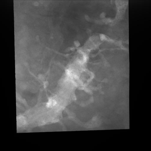
Single tilt image (zero degree tilt) through a 4 um thick section of spiny dendrite from a medium spiny neuron that was injected with Lucifer Yellow then photooxidized
Full resolution image description
tar file contains the original tilt images compressed using the compress command; osaka4.????.f.Z. Unfortunately; the original fiducial mark file was not saved.
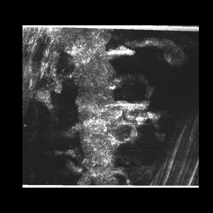
Animation description
Aligned and cropped tilt images from a striatal medium spiny neuron that was injected with Lucifer Yellow and then photooxidized. No fiducial marks were available and so the alignment and subsequent reconstruction are poor. The quality of the microscopic images is fine though, so they can be re-aligned.