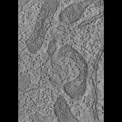
Single computed slice through a tomographic reconstruction of mitochondria from Drosophila S2 cells infected with flock house virus (FHV-1). To see the relationship between the 2D tilt image and the reconstructed area, please view the image map. Note that the reconstruction (inset on image map) is flipped vertically relative to the original tilt data.
Full resolution image description
Zip file (FHV2_vol.zip) containing a tomographic reconstruction of mitochondria from Drosophila S2 cells infected with flock house virus (FHV-1). Both the .img and .hdr files are included (fhv2.sub.img/hdr).
File format
MRC
Volume_dimension
1800, 2560, 405
Volume scale
0.0022, 0.0022, 0.0022
Animation description
Animation through the computed slices of a tomographic reconstruction of mitochondria from Drosophila S2 cells infected with flock house virus (FHV-1).
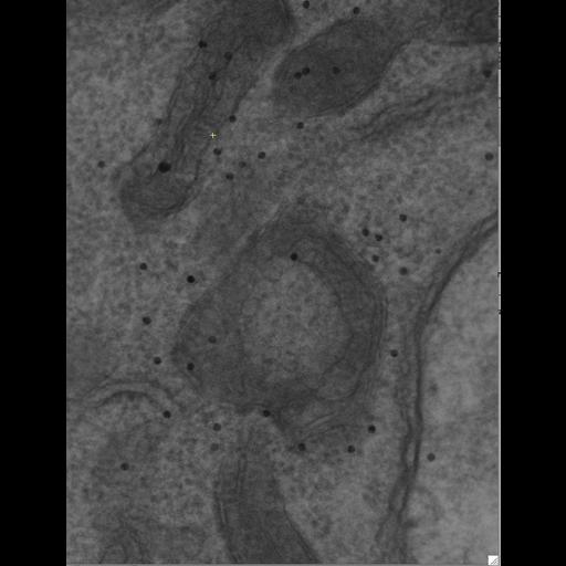
Single tilt image of a 250 um thick section of a Drosophila S2 cell infected with flock house virus (FHV-1) imaged with intermediate voltage electron microscopy, taken at zero degree tilt. The dark specks are colloidal gold particles that have been applied to the surfaces of the section to serve as fiducial marks for subsequent alignment.
Full resolution image description
Zip file containing the single axis tilt images (fhv2.st) in IMOD MRC format along with many of the ancillary files generated by iMOD.
Manual segmentation of mitochondrial membranes and replication centers (RC's) of FHV-1 using Xvoxtrace v2.13. Contours were sufaced using Synu.
Segmentation file description
Zip file containing original trace files (FHVmito2.trace and FHVmitonew.trace), along with some of the surface files in Synu (*.synu) and open inventor (*.iv) format. It appears that only some of the surface renderings for the rc objects are available; the rest will have to be regenerated; no surfaced objects associated with mitochondrial membranes were submitted. An animation of the segmented mitochondrion and the viral spherules in Quicktime format is also included (6659_seg.mov). Please note that Jinx (http://ncmir.ucsd.edu/downloads/jinx.shtm) can be used to read traces generated with Xvoxtrace. Jinx requires the original reconstruction file, available from the "Reconstruction' download page.
3D analysis of mitochondria infected with flock house virus
Description
Examination of isolated mitochondria from Drosophila following infection with flock house virus (FHV-1).
Funding agency
National Institutes of Health
Leader(s)
Guy Perkins
Collaborator(s)
Paul Ahlquist
Mark H. Ellisman
Benjamin G. Kopek
Start date
06-01-2003
End date
unspecified
Experiment
Experiment ID
6650
Title
Tomographic reconstructions of mitochondria infected with Flock House virus
Purpose
To determine structural alterations in mitochondria associated with flock house virus infection
Experimenter(s)
Guy Perkins
Microscopy product
Microscopy product ID
6659
Instrument
JEM-4000EX IVEM
Microscopy type
IVEM
Product type
SINGLE TILT
Image basename
FHV2
Spatial Axis
Image Size
Pixel Size
X
1960px
0.55 nm/pixels
Y
2560px
0.55 nm/pixels
Subject
Species
fruitfly
Scientific name
Drosophila melanogaster
Strain
melanogaster
Group by
Infection with FHV-1
Treatment
Transfection of Drosophila S2 cells with flock house virus (FHV-1); cells were harvested 12 hr post infection
Age class
not applicable
Tissue section
Microtome
Ultramicrotome
Thickness
0.25 µm
Specimen description
Structure
mitochondrion
Cell type
Drosophila S2
Imaging parameters
Type
Electron microscopy product
Recording medium
Slow scan cooled 2K CCD camera
Magification
40000
Accelerating voltage
400 KeV
Specimen preparation
Protocol used
Mitochondria were isolated from Drosophila cells as described by Echalier [77]. For electron tomography, cells were fixed in 2% parafomaldehyde and 2.5% glutaraldehyde in 0.1 M sodium cacodylate, pH 7.4, post-fixed in 1% osmium tetroxide with 0.8% potassium ferrocyanide in sodium cacodylate buffer, stained with 2% uranyl acetate, dehydrated in a graded series of ethanol, and embedded in Durcupan ACM resin. Sections were cut with a thickness of approximately 250 nm from blocks exhibiting well-preserved ultrastructure. These sections were stained for 30 min in 2% aqueous uranyl acetate, followed by 30 min in lead salts. Fiducial cues consisting of 20-nm colloidal gold particles were deposited on both sides of the section.
Imaging product type
Type
Single tilt
Description
singlet_desc
Min range
-60 degrees
Max range
60 degrees
Tilt increment
2 degrees
Citation Information
Guy Perkins, Paul Ahlquist, Mark H. Ellisman, Benjamin G. Kopek (2003) CCDB:6659, Drosophila melanogaster, mitochondrion, Drosophila S2. CIL. Dataset. https://doi.org/doi:10.7295/W9CCDB6659