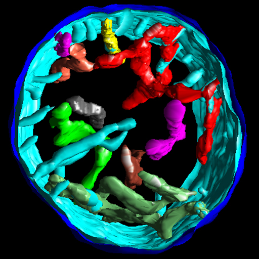
In situ structures of mitochondria in rods and cones
Leader(s)
Don Fox
University of Houston
Collaborator(s)
Guy Perkins
Start date
09-01-2001
End date
09-01-2001
Experiment
Experiment ID
33
Experiment date
07-01-2001
Title
Cone and rod mitochondria: electron tomography
Purpose
electron tomography of cone mitochondria
Experimenter(s)
Guy Perkins
Microscopy product
Microscopy product ID
54
Instrument
JEOL4000EX
Microscopy type
IVEM
Product type
single tilt
Image basename
cone1
Spatial Axis
Image Size
Pixel Size
X
1024px
0.00224 µm
Y
1024px
0.00224 µm
Subject
Species
mouse
Scientific name
mus musculus
Strain
C57BL/6
Treatment
none
Age
21 days
Age class
young adult
Tissue section
Anatomical location
retina
Microtome
ultramicrotome
Tissue product storage
Fox WT Durcupan400
Thickness
300 nm
Specimen description
Organ
eye
System
central nervous system
Structure
mitochondrion
Cell type
photoreceptor/cone
Imaging parameters
Type
Electron microscopy product
Recording medium
film
Magification
40000
Accelerating voltage
400 KeV
Imaging product type
Type
Single tilt
Description
singlet_desc
Min range
-60 degrees
Max range
60 degrees
Tilt increment
2 degrees
Citation Information
Don Fox, University of Houston, Guy Perkins (2001) CCDB:54, mus musculus, mitochondrion, photoreceptor/cone. CIL. Dataset. https://doi.org/doi:10.7295/W9CCDB54