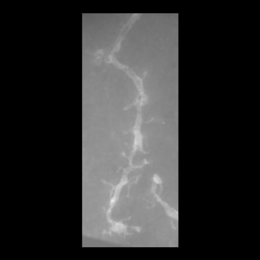
Maximum intensity project of a tomographic reconstruction of a spiny dendrite from a 4 ?m thick section throuh medium spiny neuron of mouse caudateputamen
Full resolution image description
Reconstruction of selectively stained spiny dendrite from single axis tilt tomography. A .tar file containing the reconstructed volume in both IMOD's mrc file format and in Analyze's file format.
Volume_dimension
321, 805, 151
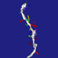
Animation description
maximum intensity projection of selectively stained spiny dendrite rotated along the y axis
Spine necks were manually defined using Analyze image edit functions. Segmentation was then performed using morphology and object definition tools provided by Analyze; segmented objects are contained in the Analyze .obj file
Experiment #5DAT KO mouse 04/22/03Description: Photoconverted dye-filled striatal medium spiny neurons for EMAnimal Info:ID#wt3wt4 Weight: 34g32g DOB: 9/30/029/30/02Protocol1. Perfusion (at Duke) Nembutal; 4% paraformaldehyde + 0.1% gluteraldehyde2. Sectioned on Vibratome (at NCMIR)Thickness = 100 micronsStore in 1X PBS in fridge3.Fill cells with Lucifer yellow4.Store slices with filled cells in 4% para in fridge5.Wash 6x with PBS 1X (on ice)6. When ready to begin photoconversion, turn on the chiller in confocal room. Set at ~4?C. The refrigerator unit should be set at TEMP < 45?C. Switch ON. Stage needs around 20 minutes to come to temperature. Pull unit out into hallway (to avoid increase in temperature).6.Place slices in 2% glut/PBS on ice for 15 minutes0.8 ml 25% gluteraldehyde2 ml 5x PBS6.2ml ddH207.Briefly wash slices in PBS8.Place slices in PBS/glycine for a few minutes38 mg glycine10 ml 1x PBS9.Follow instructions for Photoconversion of Lucifer Yellow-filled cells10. After photoconversion, remove DAB solution and wash slice 3x 10 minutes in generous volumes of PBS on ice. Must remove all DAB before beginning osmification.Microwaving protocol for osmication, dehydration, and embedding of photoconverted slices*Prepare Resin mix and let it sit covered and undisturbed until needed (instructions by fume hood in embedding area).*Rinse slices with a generous amount of cold 1X PBS on ice for ~ 10 min.*Turn on circulating bath (over 20?C, ~ RT): water bath (left hand side) will fill. *Insert temperature probe*Fill other T-beaker with water*Set temperature to 35?C*Open new bottle of 100% ethanol and prepare following dilutions:90% ethanol70% ethanol50% ethanol*Make up osmium solution under fume hood and chill on ice*1% osmium tetroxide in PBS on ice.2.0 ml PBS 5Xthen 5.5 2x distilled H2O2.5 ml Osmium 4%*Rinse w/ 2x distilled H2O ? 3 x 5min*Warm up microwave for 2 minutes on high*Label tubes & place in rack on ice*Fill tubes with osmium solution (w/ meniscus at 0.5)*Using glass hooks, transfer slices to tubes*Remove temperature probe & set temp above 50?C.*Put rack w. tubes in for 40 sec at full power*Change rear water load in T-beaker*Change osmium solution on ice and microwave for another 40 seconds at full power*Rinse samples for 2 minutes in distilled water on benchtop (at RT)*Insert petri bath with H2O under rack*Dehydration steps (2 x 40 seconds per step; all @ 35?C)1st2nd50% EtOH70% EtOH90% EtOH100% EtOH100% Acetone*All of the dehydration steps should be carried out in microcentrifuge tubes filled with 600 ml of solution. Temperature probe should be in petri dish and set for 35. Change water in rear water load when warm to touch.*Change from water to acetone in petri bath under rack ? check acetone bath level every 3 minutes*Infiltration steps (both @ 50?C): With a 50/50 mixture of resin and acetone:1 x 15 min1:1 Resin:acetone* Check rear water load at 7.5 minutesSwitch to 100% resin for 3 x 10 minutes:1st2nd3rd100% Resin*Periodically check rear water load*Flat embed samples between mould release slides and place in embedding oven under vacuum.
Imaging product type
Type
Single tilt
Description
singlet_desc
Imaging product type
Type
Through focus
Description
transmitted light z series through photoconverted medium spiny neuron
Z step
.25 microns
Psf file
061603_2
Notes
WT2 grid 16 tomo 8
Citation Information
Diana Price, Aki Laakso, Michele Cyr, Maryann Martone, Naoko Yamada, Andrea Thor, Monica Berlanga (2003) CCDB:32, mus musculus, spiny dendrite, medium spiny neuron. CIL. Dataset. https://doi.org/doi:10.7295/W9CCDB32