Alternate header for print version


Image 2D
Display image description
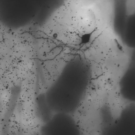
Summed Z projection through a through-focus series of a medium spiny neuron from the neostriatum of a dopamine-transporter knock out mouse, filled with Lucifer Yellow and then photooxidized. The neuron is not completely contained within the section so part of it is missing.
Full resolution image description
Zip file containing through focus series in BioRad PIC and multimage TIFF formats
Animation description
Animation stepping through the slices of the through-focus series of a medium spiny neuron from the neostriatum of a dopamine-transporter knock out mouse, filled with Lucifer Yellow and then photooxidized. The neuron is not completely contained within the section so part of it is missing. This movie was downsampled from the original file for display purposes.

Segmentation
Display image description
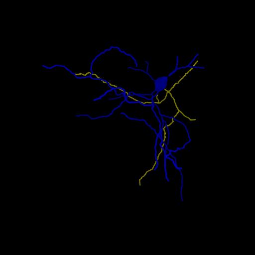
Manual tracing of dendrites using Neurolucida. Spines were traced but these were difficult to see, so the number may not be accurate.
Segmentation file description
Zip file containing Neurolucida trace file in ascii format (*_finaltrace.ASC), along with the output in VRML format. Summary files of measurements generated by Neuroexplorer for each of the parts traced are also included.

License
Attribution Only: This image is licensed under a Creative Commons Attribution License. View License Deed | View Legal Code

CCDB:3659*  Cite 
Project: P1207
Project name
Correlative microscopic characterization of dendritic spines in a transgenic mouse model of hyperdopaminergia: The dopamine transporter knockout mouse
Description
Multiscale characterization of DAT KO transgenic mouse
Funding agency
NIH
Leader(s)
Diana Price
Collaborator(s)
Aki Laakso
Michele Cyr
Maryann Martone
Naoko Yamada
Andrea Thor
Monica Berlanga
Start date
01-01-2003
End date
unspecified
 
Experiment
Experiment ID
20
Experiment date
01-09-2003
Title
P1207 Experiment 1
Purpose
EMT reconstructions of medium spiny neuron dendrites
Experimenter(s)
Diana Price
Masako Terada
Andrea Thor
Microscopy product
Microscopy product ID
3659
Instrument
Biorad 1024 MRC confocal
Microscopy type
laser scanning confocal
Product type
THROUGH FOCUS SERIES
Image basename
DATKOA
Spatial Axis Image Size Pixel Size
X 1024px 0.158 um/pixels
Y 1024px 0.158 um/pixels
Subject
Species
mouse
Scientific name
mus musculus
Strain
C57BL/129SvJ
Group by
genetic manipulation
Treatment
Knockout of dopamine transporter
Age
6 months
Age class
adult
Tissue section
Anatomical location
neostriatum DATKOA
Microtome
Vibratome
Tissue product storage
DAT#1
Thickness
100 µm
Specimen description
Organ
brain
System
central nervous
Map location
View location
Cell type
medium spiny neuron
Atlas coordinate
0, 0, 0,
Imaging parameters
Type
Light microscopy product
Immersion medium
oil
Mounting medium
resin
Lens
Zeiss Plan APOCHROMAT
Lens magnification
X
Numerical aperture
1.4
Notes
mmartone
Imaging product type
Type
Through focus
Z step
.54 um