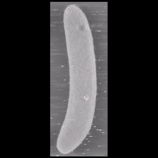
A maximum intensity projection image of a caulobacter crescentus specimen taken on an IVEM.
Full resolution image description
A .tar file containing to .mrc format slice by slice volume
reconstructions. One is a trimmed stack of the other larger volume. Both
files were made using IMOD's tomographic reconstruction program.
Volume_dimension
1156, 3116, 1269
Animation description
A .mpg file movie of the slice by slice reconstructed volume of a caulobacter crescentus specimen.
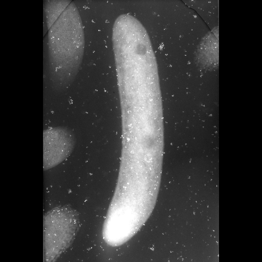
A Caulobacter crescentus specimen taken at a 0 degree tilt. Gold fiducials are 10 and 15 nm in diameter.
Full resolution image description
A .tar file containing all processing files for an IMOD volume
reconstruction. Along with necessary .st, .preali, .ali and .rawtlt files
is a notes.txt file containing some information necessary for proper
processing.
Animation description
a .mpg format movie file showing the aligned tilt series taken on an IVEM, shot onto film.
To produce a series of tomographic reconstructions of the normal morphology of caulobacter using traditional fixing methods.
Experimenter(s)
Tom Deerinck
Guido Gaietta
Microscopy product
Microscopy product ID
3699
Instrument
JEOL 4000 #1
Microscopy type
IVEM
Product type
SINGLE TILT
Image basename
cb0
Spatial Axis
Image Size
Pixel Size
X
4482px
Y
6678px
Subject
Species
Caulobacter crescentus
Scientific name
Caulobacter crescentus (Snc04)
Strain
Snc04
Age class
N/A
Tissue section
Anatomical location
N/A
Tissue product storage
N/A
Thickness
0.4 µm
Specimen description
Cell type
Caulobacter crescentus
Imaging parameters
Type
Electron microscopy product
Recording medium
film
Magification
30000
Specimen preparation
Protocol used
Induced for 1 hour at 30 degrees CLabeled in M2G medium for 2 hours at 30 degrees C (in the presence of 1.8 micromolar ReAsH-EDT2) and photoconvertedSamples were fixed with 2.5% glutaraldehyde with 1% acrolein in 0.05M sodium cacodylate pH 7.4 with 3 mM CaCl2 and 1 % sucrose followed by conventional osmium tetroxide/potassium ferrocyanide post-fixation, dehydration and embedding.
Imaging product type
Type
Single tilt
Description
singlet_desc
Min range
-60 degrees
Max range
60 degrees
Tilt increment
2 degrees
Notes
10 and 15 nm gold fiducials
Citation Information
Lucy Shapiro, Harley McAdams, Patrick H. Viollier, Tom Deerinck, Mason Mackey, John Crum, Mark Ellisman (2003) CCDB:3699, Caulobacter crescentus (Snc04), Caulobacter crescentus. CIL. Dataset. https://doi.org/doi:10.7295/W9CCDB3699