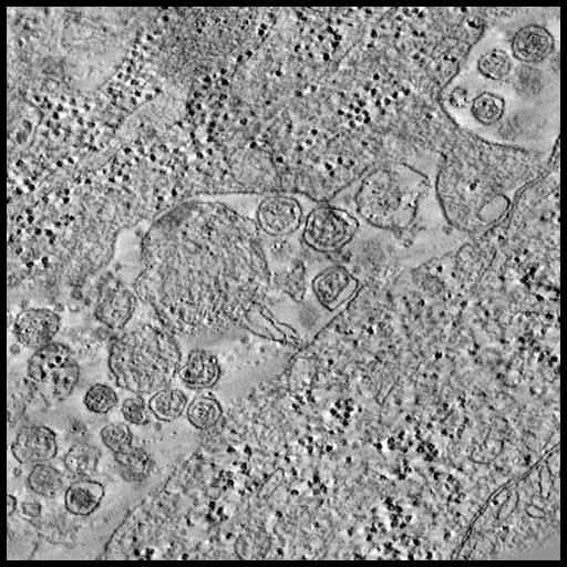
Serial tomographic reconstruction from four serial tomograms showing a large number of HTLV-1 particles in the space between a chronically infected MS9 cell (top) and a potential Jurkat cell (bottom). Viral particles are characterized by an electron-dense core surrounded by a less dense area and bounded by a viral membrane.
Full resolution image description
Serial tomographic reconstruction from four serial tomograms in MRC format. Filename = map_3950.ccp4
Volume_dimension
1603, 1058, 529
Volume scale
0.00121, 0.00121, 0.00121
Animation description
Animation through the computed slices of a serial tomographic reconstruction from four serial tomograms showing a large number of HTLV-1 particles in the space between a chronically infected MS9 cell (top) and a potential Jurkat cell (bottom). Viral particles are characterized by an electron-dense core surrounded by a less dense area and bounded by a viral membrane.
Electron tomography of the HTLV-1 virological synapse
Description
Tomography of the virological synapse formed between an HTLV-1 infected cells and target cells.
Funding agency
Wellcome Trust
Leader(s)
Endre Majorovits
Collaborator(s)
Charles Bangham
Stephen Fuller
Mohamed Nejmeddine
Start date
02-02-2004
End date
02-02-2004
Experiment
Experiment ID
6104
Title
Tomography of the HTLV-1 virological synapse in cultured cells
Purpose
Tomographic reconstruction of virological synapse formed between a chronically infected cell line (MS9) and Jurkat cells
Experimenter(s)
Endre Majorovits
Mohamed Nejmeddine
Microscopy product
Microscopy product ID
3950
Instrument
FEI Tecnai F30 FEG
Microscopy type
TEM
Product type
SINGLE TILT
Image basename
HTLV1_VS_MS9_virus
Spatial Axis
Image Size
Pixel Size
X
1312px
3.14 nm/pixels
Y
643px
3.14 nm/pixels
Subject
Species
human
Scientific name
homo sapiens
Strain
sapiens
Group by
viral infection
Treatment
infection by HTLV-1, molecular clone pHTLV-X1MT
Age class
n/a
Tissue section
Microtome
ultramicrotome
Thickness
0.3 µm
Specimen description
System
blood
Structure
virological synapse
Cell type
MS9
Imaging parameters
Type
Electron microscopy product
Recording medium
Slow scan cooled 2K CCD camera
Magification
10
Accelerating voltage
300 KeV
Notes
These data were recorded in the Laboratory for 3D Electron Microscopy of Cells in Boulder, Colorado with a FEI Tecnai F30 FEG-TEM without energy filter.
Specimen preparation
Protocol used
The HTLV-1- immortalized cell line, MS9 was a gift from Dr. David Derse, National Cancer Institute, Maryland, USA. MS9 cells were derived by co-culture of phorbol-12- myristate-13- acetate-activated human peripheral PBMCs with DBS-FRhL (clone B5) cells that were infected with the HTLV-1 molecular clone, pHTLV-X1MT. MS9 cells were cultured in RPMI 1640 (Sigma-Aldrich Company Ltd, Dorset, UK) supplemented with 2 mM glutamine (Invitrogen Ltd, Paisley, UK), 100 IU/ml penicillin (Invitrogen Ltd, Paisley, UK), 100 IU/ml streptomycin (Invitrogen Ltd, Paisley, UK), 20% heat-inactivated fetal calf serum (PAA Laboratories Ltd, Somerset, UK) and 100 U/ml recombinant interleukin 2 (IL-2; Sigma-Aldrich Company Ltd, Dorset, UK). Jurkat cells (clone E6.1) were obtained from ATCC, Middlesex, UK. Jurkat E6.1 is a clone of the Jurkat-FHCRC cell line, a derivative of the Jurkat cell line [32]. Before processing for EM and/or IFM the cells were mixed 1:1 with Jurkat cells and incubated for 30 minutes at 37uC and 5% CO2 to induce cell-cell conjugates.Samples were prepared with different buffers and different amounts of fixatives, depending on whether they were destined to be immunostained with the mAb anti-Gag p19 (GIN7) or to be stained with magnesium uranyl acetate (Sigma-Aldrich Company Ltd, Dorset, UK). In general, the fixation of the samples was performed sequentially in two distinct fixative solutions: Solution A, 2% paraformaldehyde (PFA) (Electron Microscopy Science, Hatfield, PA, USA) and 0.5% glutaraldehyde (Agar Scientific Ltd, Essex, UK) in sodium cacodylate buffer 0.1 M, pH 7.2 (Sigma-Aldrich Ltd, Dorset, UK); and Solution B, 1% osmium tetroxide (OsO4) in PBS (Sigma-Aldrich Ltd, Dorset, UK).After incubating the samples to induce cell-cell conjugates, they were pre-fixed in the 10X diluted PFA/glutaraldehyde (Solution A) for 10 minutes before centrifugation. After centrifuging for 5 minutes at 1000 g the samples were fixed for another hour with the undiluted fixative (Solution A). The samples were washed 3 times either with PBS 1% BSA or with sodium cacodylate buffer 0.1 M, pH 7.2, and then processed for antibody staining as described below or post-fixed with 1% OsO4 (Solution B) and stained in magnesium uranyl acetate overnight, respectively.The fixed samples were dehydrated through a series of ethanol exchanges and embedded in Agar 100 resin (Agar Scientific Ltd., Stansted, UK). The samples were baked at 60uC overnight to give solid blocks that were sectioned with an ultramicrotome (Leica Ultracut UCT, Leica Microsystems GmbH, Wetzlar, Germany) at a thickness of 60 to 300 nm. Thin samples (60 to 100 nm) were floated onto 200/300 mesh square Ni grids (Agar Scientific Ltd., Stansted, UK). Thick samples for tomography (200 to 300 nm) were either floated onto 200 mesh square Ni grids or onto formvar film coatedCu 162 mm slot grids (Agar Scientific Ltd., Stansted, UK) to allow for the collection of serial sections. Samples that were not antibody labelled were stained with lead citrate (Leica Ultrostain2) for 3 to 10 minutes, depending on the thickness of the sections, by floating the grids on drops of the staining solution. For electron tomography the grids were covered with 10 nm or 15 nm fiducial gold beads (Sigma-Aldrich Company Ltd, Dorset, UK) to facilitate image processing.
Imaging product type
Type
Single tilt
Description
singlet_desc
Min range
-60 degrees
Max range
60 degrees
Tilt increment
1 degrees
Notes
Four serial tomograms were obtained
Citation Information
Endre Majorovits, Charles Bangham, Stephen Fuller, Mohamed Nejmeddine (2004) CCDB:3950, homo sapiens, virological synapse, MS9. CIL. Dataset. https://doi.org/doi:10.7295/W9CCDB3950