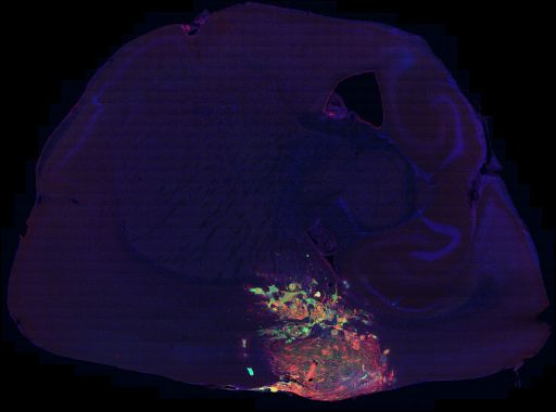
Processed mosaic of a sagittal section through the brain of mouse model of glioma. To produce transformed cells, creGFAP mice were injected in the cortex with Ras/s.i.p53 virus and perfused after 5 weeks. In this system, GFP is introduced by the virus and expression is induced by an IRES site in the mRNA. Thus, the GFP gene is expressed whenever the virus infects a cell expressing GFAP (green). The section was immunolabeled for nestin (red) and counterstained with DAPI (blue). See MP:7857 for a section from the same brain labeled with GFAP.
To characterize glial morphology and molecular markers in a mouse model of glioblastoma
Funding agency
NIH
Leader(s)
Inder Verma
Collaborator(s)
Dinorah Friedmann-Morvinski
Eric Bushong
Mark Ellisman
Start date
06-01-2007
End date
unspecified
Experiment
Experiment ID
7596
Title
Batch 14
Purpose
Three cre-GFAP mice were injected in the cortex with Ras / s.i. p53 virus. The mice were perfused 5 weeks after injection.
Experimenter(s)
Dinorah Friedmann-Morvinski / Eric Bushong
Microscopy product
Microscopy product ID
8063
Instrument
Nikon RTS 2000 multiphoton microscope
Microscopy type
MULTIPHOTON
Product type
MOSAIC
Image basename
BT14BR1_NESTIN-1.IMG
Spatial Axis
Image Size
Pixel Size
X
512px
0.36 um/pixels
Y
512px
0.36 um/pixels
Y
11px
Subject
Species
mouse
Scientific name
mus musculus
Strain
C57Bl/6
Treatment
All animals received the same treatment.
creGFAP mice were injected in the cortex with Ras/s.i.p53 virus and perfused after 5 weeks.
Age class
adult
Tissue section
Anatomical location
brain
Microtome
vibratome
Thickness
70 µm
Specimen description
Organ
brain
System
central nervous system
Imaging parameters
Type
Light microscopy product
Immersion medium
oil
Mounting medium
gelvatol
Lens
Nikon 40X oil
Lens magnification
x
Numerical aperture
1.30
Notes
ebushong
Specimen preparation
Protocol used
A slice was taken from cryoprotectant and washed for 3x 10 minutes in PBS.The slice was placed in blocking buffer at room temp for 1 hour.Blocking buffer: PBS, 3% NDS, 1% BSA, 1% CWFG, 0.3% Triton X-100The slice was briefly washed in working buffer.Working buffer: 1 in 10 dilution of blocking buffer in PBS, plus 0.1% tritonThe slice was placed in working buffer containing 1:500 dilution of chicken anti-nestin antibody (Neuromics Cat#CH23001, Lot#400588). The slice was left in cold room overnight.The slice was washed 3x 20 mintues in working buffer at room temp.The slice was placed in working buffer containing 1:200 dilution of donkey anti-chicken RRX antibody. The slice was left in cold room overnight.The slice was washed 1X 15 minutes in PBS.The slice was placed in DAPI/PBS solution for 15 minutes.The slice was washed 2x 15 minutes in PBS.The slice was coverslipped in Gelvatol and stored in fridge until imaged.
Imaging product type
Type
Mosaic
Description
nestin labeling of creGFAP brain 5 weeks post inj with Ras/s.i.p53 virus into cortex
X position
46 tiles
Y position
36 tiles
Citation Information
Inder Verma, Dinorah Friedmann-Morvinski, Eric Bushong, Mark Ellisman (2007) CCDB:8063, mus musculus. CIL. Dataset. https://doi.org/doi:10.7295/W9CCDB8063