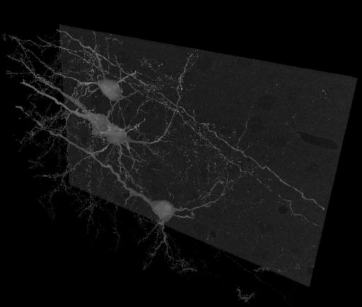
Golgi impregnated pyramidal neurons in the neocortex of an adult mouse. Contrast is reversed so that Golgi stain appears bright against the unstained background. A maximum intensity projection of the dataset is available in the thumbnail and by clicking the JPEG tab.
Full resolution image description
Full resolution file in IMOD format. The raw series had 1560 planes total, but there is no information available as to why some planes were left off the reconstruction.
Downsampled image description
Volume binned by a factor of 2 in the x, y and z planes: 2048 X 2048 X 632 slices
File format
MRC
Volume_dimension
4096, 4096, 1266
Volume scale
0.047, 0.047, 0.06
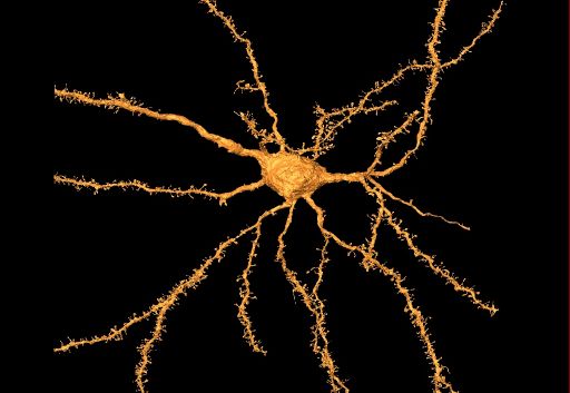
Animation description
Animation showing the segmentation of individual neurons from a reconstruction of Golgi-impregnated mouse neocortex using serial block face SEM. Segmentation files are available for download under the segmentation section.
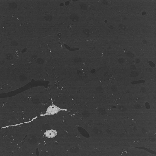
Single slice through Golgi-impregnated neurons from the mouse neocortex imaged using serial block face SEM. Contrast is reversed so that electron dense regions appear bright.
Full resolution image description
Tar file containing raw data files in dm3 format
Animation description
Slice by slice animation of Golgi-impregnated neurons from the mouse neocortex imaged using serial block face SEM. Contrast is reversed so that electron dense regions appear bright.
The Whole Brain Catalog™ is a ground-breaking, open-source, 3-D virtual environment developed by a team of researchers from UC San Diego under the Whole Brain Project™. The Catalog aims to connect members of the international neuroscience community to facilitate solutions for today's intractable challenges in brain research through cooperation and crowd sourcing. CCDB provides the backend services for very large scale image data sets on mouse brain derived from high resolution light and electron microscopy
Funding agency
Ted Waitt Family Foundation
Leader(s)
Mark Ellisman
Stephen Larson
Collaborator(s)
Sarah Maynard
Eric Bushong
Maryann Martone
Start date
08-05-2009
End date
unspecified
Experiment
Experiment ID
8238
Title
Identified neuron reconstructions using SBFSEM
Purpose
To prepare specimens with suitable contrast for whole cell reconstruction of stained cells using serial block face SEM
Perfused with 4% PFA, 0.1% glut. Postfixed 1 hour in same fix on ice. Left hemisphere cut into 2 mm thick coronal slabs. Tissue placed into 2.3% potassium dichromate / 0.4% OsO4 for 3 days at room temp. Tissue blotted with filter paper and then placed in 0.7% silver nitrate in dark for 3 days. Tissue was dehydrated, infiltrated and embedded in Durcupan in Beem capsules.
Imaging product type
Type
Serial section
Description
Serial block face imaging
Z resolution
60 nm
Citation Information
Mark Ellisman, Stephen Larson, Sarah Maynard, Eric Bushong, Maryann Martone (2009) CCDB:8244, mus musculus, neocortex pyramidal neuron. CIL. Dataset. https://doi.org/doi:10.7295/W9CCDB8244