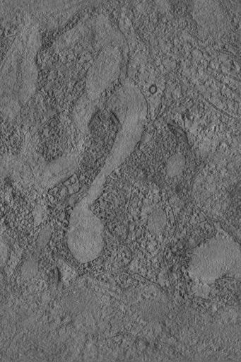
Double tilt electron tomographic reconstruction of a glial process apposing two synapses in the neuropil of the stratum radiatum of hippocampal area CA1 of the adult rat.
Full resolution image description
Double tilt electron tomographic volume of astrocytic processes in the stratum radiatum of hippocampal area CA1 in TIFF format (e1.tiff).
File format
TIFF
Volume_dimension
1000, 1500, 140
Volume scale
0.0011, 0.0011, 0.0012
Animation description
Animation through the computed slices of a double tile tomographic reconstruction of a glial process apposing two synapses in the neuropil of the stratum radiatum of hippocampal area CA1 of the adult rat.
Electron tomographic analysis of synaptic structure in the adult rat cortex
Description
Novel specimen preparation techniques were combined with electron tomography to provide new views of synaptic structure in the cortex of the adult rat
Leader(s)
Alain C. Burette
Collaborator(s)
Richard Weinberg
John Crum
Mark Ellisman
Maryann Martone
Start date
09-01-2000
End date
09-01-2000
Experiment
Experiment ID
8475
Title
Tomographic imaging of synapses
Purpose
Imaging of synapses prepared with an osmium free aldehyde fixation protocol
Experimenter(s)
Alain C. Burette
Microscopy product
Microscopy product ID
8638
Instrument
JEOL4000EX
Microscopy type
IVEM
Product type
DOUBLE TILT
Image basename
e1
Subject
Species
rat
Scientific name
rattus rattus
Strain
Sprague Dawley
Age class
adult
Tissue section
Anatomical location
Hippocampal area CA1 or cortical area S1
Thickness
0.12 µm
Specimen description
Organ
Brain
System
central nervous system
Structure
neuropil
Imaging parameters
Type
Electron microscopy product
Recording medium
4K CCD
Magification
0
Accelerating voltage
400 KeV
Specimen preparation
Protocol used
Adult male Sprague-Dawley rats (250-500 g) were deeply anesthetized with pentobarbital (60 mg/kg, IP) and sacrificed by intra-aortic perfusion with 2% glutaraldehyde and 2% freshly-depolymerized paraformaldehyde in 0.1 M phosphate buffer (PB, pH 7.4), after a brief flush with heparinized saline. Blocks of fixed forebrain were sectioned on a Vibratome at 50 um, collected and stored in PB at 4 degrees C. Sections containing regions of interest were prepared for electron microscopy according to modifications of the protocol described in Phend et al. 1995. In 1% uranyl acetate. Additional metal salts were tested, including potassium ferrocyanide, chromium potassium sulfate, osmium trichloride, iridium tetrabromide, and mercuric acetate. Particularly fine grain and visualization of structure was seen by combining uranyl acetate with 0.1% PtCl4; for that reason, the present manuscript is based on observations from this material.Sections collected in glass vials on a shaker at 4 degrees C were incubated 40 min in 1% tannic acid (Mallinkrodt) in 0.1M maleate buffer pH 6.0 (MB), then 20 min in 0.1% CaCl2 in MB, then 40 min in a mixture of 1% uranyl acetate (Electron Microscopy Sciences) and 0.1% PtCl4 (Pfaltz & Bauer), then rinsed in MB. Sections were then dehydrated through graded ethanol solutions into propylene oxide. Sections were infiltrated with Epon-Spurr resin at room temperature, sandwiched between two sheets of Aclar plastic, and heat-polymerized at 60 degrees C. Chips of S1 cortex and CA1 hippocampus were glued to plastic blocks and thin sections cut on an ultramicrotome with a diamond knife. For electron tomography, ~120 nm sections were collected on 100 mesh hexagonal gold grids. To test retention of antigenicity, postembedding immunocytochemistry was performed: grids were immunoreacted with NR2A/B primary antibody (Chemicon, AB1548, lot# 0509010940) and visualized with 20 nm gold particles, as described (Phend et al., 1992).
Imaging product type
Type
Double tilt
X min range
-70 degrees
X max range
70 degrees
X tilt increment
2 degrees
Y min range
-70 degrees
Y max range
70 degrees
Y tilt increment
2 degrees
Citation Information
Alain C. Burette, Richard Weinberg, John Crum, Mark Ellisman, Maryann Martone (2000) CCDB:8638, rattus rattus, neuropil. CIL. Dataset. https://doi.org/doi:10.7295/W9CCDB8638