Alternate header for print version


Licensing
Attribution Non-Commercial; No Derivatives:This image is licensed under a Creative Commons Attribution, Non-Commercial, No Derivatives License. View License Deed | View Legal Code
tweet  
*CIL – Cell Image Library accession number. Please use this to reference an image.

CIL:10974*  Cite 
Description

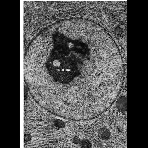
Transmission electron micrograph illustrating the 'classic' appearance of the nucleus in osmium tetroxide fixed material. In this pancreatic acinar cell nucleus, the only prominent feature is the large nucleolus. Figure 112 from Chapter 4 (Nucleus) of 'The Cell, 2nd Ed.' by Don W. Fawcett M.D. A PDF copy of the corresponding chapter is available on the ASCB's BioEDUCATE website.

Attribution
Names
Don W. Fawcett
Published
ISBN 0721635849
Link
ASCB BioEDUCATE Program
OTHER
from Don W. Fawcett M.D ‘The Cell’ 2nd edition, W.B. Saunders Chapter 4 (Nucleus)
Citation
Digital Object Identifier (DOI)
doi:10.7295/W9CIL10974
Archival Resource Key (ARK)
ark:/b7295/w9cil10974
Grouping This image is part of a group.
Imaging
Image Type
film
Image Mode
transmission electron microscopy (TEM)
detection of electrons
Parameters Imaged
electron density
Source of Contrast
differences in adsorption or binding of stain
Visualization Methods
uranyl salt
osmium tetroxide
lead salt
Processing History
Image extracted from .pdf file of 'The Cell'
Data Qualifiers
processed data
Sample Preparation
Methods
tissue in plastic embedment
osmium tetroxide fixed tissue
Relation To Intact Cell
microtome-sectioned tissue
Dimensions
Spatial Axis Image Size Pixel Size
X 914px ——
Y 1260px ——