Alternate header for print version
Advanced search
Contributors
Help
Submit
Search
menu
Cell Process
Cell Component
Cell Type
Organism
Microbial
Alzheimer's
Data Sets
University of California, San Diego
9500 Gilman Drive
La Jolla, CA 92093-0608, USA
Voice
: (858) 534-0276
Fax
: (858) 534-7497
Email
: dorloff@ncmir.ucsd.edu
Grouped images - the images shown below are related
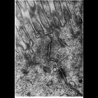
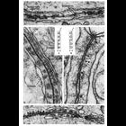
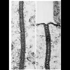
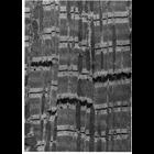
CIL:11180
NCBI Organism Classification
Rattus
Biological Process
maintenance of apical/basal cell polarity
Cellular Component
apical junction complex
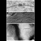
Electron micrograph of the junctional complex of intestinal epithelial cells of ...
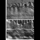
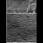
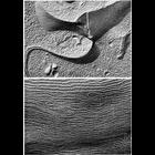
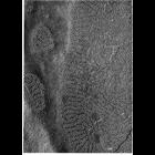
CIL:11182
NCBI Organism Classification
Bufo
Biological Process
cell junction organization
Cellular Component
tight junction
Freeze fracture replicas of zonula occludens junctions from the large intestine...
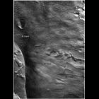
CIL:11183
NCBI Organism Classification
Xenopus laevis
Biological Process
cell junction organization
Cellular Component
tight junction
Freeze fracture replica of zonula occludens junctions from the small intestine ...
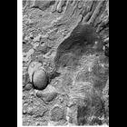
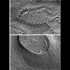
CIL:11192
NCBI Organism Classification
Cavia porcellus
Biological Process
cell junction organization
Cellular Component
tight junction
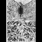
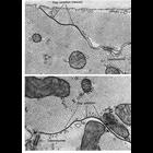
Sertoli-Sertoli occluding junctions from guinea pig testis. Figures 67 (left) a...
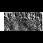
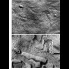
CIL:11193
NCBI Organism Classification
Rattus
Biological Process
cell junction organization
Cellular Component
occluding junction
Occluding junctions in Sertoli cells of ram and rat testis. Multiple focal site...
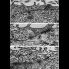
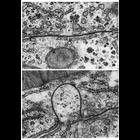
CIL:11196
NCBI Organism Classification
Rattus
Biological Process
cell-cell junction organization
Cellular Component
occluding junction
Upper panel shows a typical zonula occludens of rat intestinal epithelium, prepa...
CIL:11197
NCBI Organism Classification
Rattus
Biological Process
cell-cell adhesion
Cellular Component
occluding junction
A freeze fracture replica of a Sertoli cell junction from rat testis shows more ...
CIL:11199
NCBI Organism Classification
Rattus
Biological Process
cell-cell adhesion
Cellular Component
zonula adherens
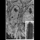
These examples of ependymal epithelium from the rat brain represent epithelia th...
CIL:11201
NCBI Organism Classification
Siphonaptera
Biological Process
cell-cell junction organization
Cellular Component
septate junction
Epithelia of invertebrates contain zona continua and septate junctions not found...
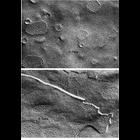
CIL:11202
NCBI Organism Classification
Rhodnius prolixus
Biological Process
cell-cell junction organization
Cellular Component
cell-cell junction
Freeze-fracture images of continuous junctions along epithelial cells from the m...
CIL:11204
NCBI Organism Classification
Siphonaptera
Biological Process
cell-cell junction organization
Cellular Component
cell-cell junction
Freeze-fracture preparation of a continuous junction on the lateral surface of e...
CIL:11206
NCBI Organism Classification
Periplaneta americana
Biological Process
cell-cell junction organization
Cellular Component
septate junction
Figures 81 (upper) and 82 (lower) from Chapter 3 (Junctional Specializations) of...
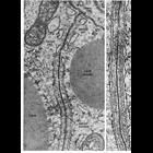
CIL:11209
NCBI Organism Classification
Phodopus
Biological Process
cell adhesion
Cellular Component
desmosome
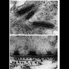
Upper panel, adjoining portions of two cells in the stratum spinosum of hamster ...
CIL:11211
NCBI Organism Classification
Opsanus tau
Biological Process
cell adhesion
Cellular Component
desmosome
Upper: capillary endothelial cell junction in the rete mirabile of the gas bladd...
CIL:11213
NCBI Organism Classification
Rattus
Biological Process
cell adhesion
Cellular Component
desmosome
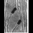
Stratified squamous epithelium from the esophagus of the rat. Keratin filaments...
CIL:11214
NCBI Organism Classification
Aphrodita aculeata
Biological Process
cell adhesion
Cellular Component
desmosome
Glial cells in the nerve cord of the annelid worm, Aphrodita aculeata are rich i...
CIL:11215
NCBI Organism Classification
Mus musculus
Biological Process
cell adhesion
Cellular Component
desmosome
Images of desmosomes from conventional EM and freeze fracture preparations show ...
CIL:11221
NCBI Organism Classification
Carassius auratus
Biological Process
cell communication
Cellular Component
gap junction
Typical organization of gap junctions as seen in thin sections (upper) and repli...
CIL:11225
NCBI Organism Classification
none specified
Biological Process
cell communication
Cellular Component
gap junction
Replica of a freeze-fractured gap junction presents the inner half-membrane on o...
CIL:11226
NCBI Organism Classification
Mus musculus
Biological Process
cell communication
Cellular Component
gap junction
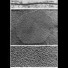
Gap junctions isolated from mouse liver and negatively stained with uranyl forma...
CIL:11230
NCBI Organism Classification
Rattus
Biological Process
cell communication
Cellular Component
gap junction
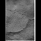
Gap junctions from a granulosa cell of a rat ovarian follicle. Variability in s...
CIL:11232
NCBI Organism Classification
none specified
Biological Process
cell communication
Cellular Component
gap junction
Gap junctions in ciliary epithelium under conditions of oxygenation (upper) and ...
CIL:11235
NCBI Organism Classification
none specified
Biological Process
cell communication
Cellular Component
gap junction
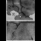
Upper: a gap junction on the boundary between two hepatic cells shows a typical ...
CIL:11237
NCBI Organism Classification
Macaca mulatta
Biological Process
cell communication
Cellular Component
gap junction
Representative examples of gap junctions from vertebrates (ciliary epithelium fr...
CIL:11243
NCBI Organism Classification
Felis catus
Biological Process
cell communication
Cellular Component
gap junction
Papillary muscle from cat heart shows a step-like end-to-end junction of two car...
CIL:11245
NCBI Organism Classification
Felis catus
Biological Process
cell communication
Cellular Component
gap junction
Gap junctions in papillary muscle from cat heart. These communicating junctions...